
Page 51 - Read Online
P. 51
Loong et al. J Transl Genet Genom 2023;7:27-49 https://dx.doi.org/10.20517/jtgg.2022.20 Page 45
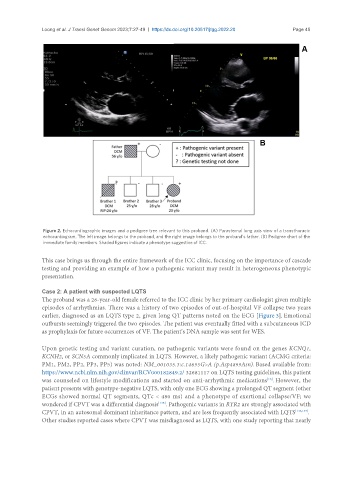
Figure 2. Echocardiographic images and a pedigree tree relevant to this proband. (A) Parasternal long axis view of a transthoracic
echocardiogram. The left image belongs to the proband, and the right image belongs to the proband’s father. (B) Pedigree chart of the
immediate family members. Shaded figures indicate a phenotype suggestive of ICC.
This case brings us through the entire framework of the ICC clinic, focusing on the importance of cascade
testing and providing an example of how a pathogenic variant may result in heterogeneous phenotypic
presentation.
Case 2: A patient with suspected LQTS
The proband was a 26-year-old female referred to the ICC clinic by her primary cardiologist given multiple
episodes of arrhythmias. There was a history of two episodes of out-of-hospital VF collapse two years
earlier, diagnosed as an LQTS type 2, given long QT patterns noted on the ECG [Figure 3]. Emotional
outbursts seemingly triggered the two episodes. The patient was eventually fitted with a subcutaneous ICD
as prophylaxis for future occurrences of VF. The patient’s DNA sample was sent for WES.
Upon genetic testing and variant curation, no pathogenic variants were found on the genes KCNQ1,
KCNH2, or SCN5A commonly implicated in LQTS. However, a likely pathogenic variant (ACMG criteria:
PM1, PM2, PP2, PP3, PP5) was noted: NM_001035.3:c.14695G>A (p.Asp4899Asn). Based available from:
https://www.ncbi.nlm.nih.gov/clinvar/RCV000182849.2/ 32681117 on LQTS testing guidelines, this patient
[16]
was counseled on lifestyle modifications and started on anti-arrhythmic medications . However, the
patient presents with genotype-negative LQTS, with only one ECG showing a prolonged QT segment (other
ECGs showed normal QT segments, QTc < 480 ms) and a phenotype of exertional collapse/VF; we
[146]
wondered if CPVT was a differential diagnosis . Pathogenic variants in RYR2 are strongly associated with
CPVT, in an autosomal dominant inheritance pattern, and are less frequently associated with LQTS [146,147] .
Other studies reported cases where CPVT was misdiagnosed as LQTS, with one study reporting that nearly