
Page 46 - Read Online
P. 46
Page 40 Loong et al. J Transl Genet Genom 2023;7:27-49 https://dx.doi.org/10.20517/jtgg.2022.20
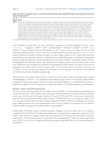
Table 1. ICC genes are routinely used in our assessment panel consisting of genes identified through our own experience and those in
Illumina Trusight panel [114]
No. of Genes
genes
176 ACTA2, ACTC1, APOB, COL3A1, DSC2, DSG2, DSP, FBN1, GLA, KCNH2, KCNQ1, LDLR, LMNA, MYBPC3, MYH11, MYH7, MYL2, MYL3,
PCSK9, PKP2, PRKAG2, RYR1, RYR2, SCN5A, SMAD3, SMAD4, TGFBR1, TGFBR2, TMEM43, TNNI3, TNNT2, TPM1, ABCC9, ABCG5,
ABCG8, ACTA1, ACTN2, AKAP9, ALMS1, ANK2, ANKRD1, APOA4, APOA5, APOC2, APOE, BAG3, BRAF, CACNA1C, CACNA2D1,
CACNB2, CALM1, CALM2, CALM3, CALR3, CASQ2, CAV3, CAVIN4, CBL, CBS, CETP, COL5A1, COL5A2, COX15, CREB3L3, CRELD1,
CRYAB, CSRP3, CTF1, DES, DMD, DNAJC19, DOLK, DPP6, DTNA, EFEMP2, ELN, EMD, EYA4, FBN2, FHL1, FHL2, FKRP, FKTN, FXN,
GAA, GATAD1, GCKR, GJA5, GPD1L, GPIHBP1, HADHA, HCN4, HFE, HRAS, HSPB8, ILK, JAG1, JPH2, JUP, KCNA5, KCND3, KCNE1,
KCNE2, KCNE3, KCNJ2, KCNJ5, KCNJ8, KLF10, KRAS, LAMA2, LAMA4, LAMP2, LDB3, LDLRAP1, LMF1, LPL, LTBP2, MAP2K1,
MAP2K2, MIB1, MYH6, MYLK, MYLK2, MYO6, MYOZ2, MYPN, NEXN, NKX2-5, NODAL, NOTCH1, NPPA, NRAS, PDLIM3, PLN,
PRDM16, PRKAR1A, PTPN11, RAF1, RANGRF, RBM20, SALL4, SCN1B, SCN2B, SCN3B, SCN4B, SCO2, SDHA, SELENON, SGCB, SGCD,
SGCG, SHOC2, SLC25A4, SLC2A10, SNTA1, SOS1, SREBF2, TAZ, TBX20, TBX3, TBX5, TCAP, TGFB2, TGFB3, TMPO, TNNC1, TRDN,
TRIM63, TRPM4, TTN, TTR, TXNRD2, VCL, ZBTB17, ZHX3, ZIC3
data (if available), among others. In silico tools used to categorize and predict implications of the variant
[116]
a
[118]
[117]
[119]
i n c l u d e Polyphen2 , SIFT , LRT , MutationTaster , Fathmm , phyloP , CADD , n d
[121]
[120]
[115]
DANN . These provide functional predictions for the variants, such as whether the amino acid
[122]
substitution affects protein structure and function. Functional data include previous in vivo or in vitro
studies published on specific variants and genes. To achieve high confidence, the studies must model the
natural disease state as closely as possible. Family history of similar phenotypes is considered in segregation
analysis, which also affects the variant classification based on the condition’s inheritance pattern. Of note,
the segregation of a particular variant with a phenotype in a family is evidence for the linkage of the variant
to the disorder but not any indication of the level of pathogenicity of the variant. Conversely, the absence of
segregation provides evidence of the benign nature of the variant unless incomplete penetrance is
considered. De novo variants are considered strong support for pathogenicity if the parents of the patients
are sufficiently evaluated clinically and genetically.
All variants are cross-referenced with other curations recorded in the literature, including disease-causing
variants flagged in ClinVar . The variants are then analyzed in the context of the patient’s clinical history
[123]
to identify (1) the disease-specific variant(s); and (2) incidental findings. To accomplish this, variants are
classified according to ACMG classifications: benign, likely benign, VUS, likely pathogenic, and pathogenic.
Genome, exome, and panel sequencing
The choice of genome sequencing (WGS), exome sequencing (WES), or panel sequencing in assessing ICC
considers factors such as the evidence surrounding gene-disease implications, cost-effectiveness, and depth
of coverage. With recent technological advances, the cost of WGS and WES has decreased significantly
without sacrificing the depth of sequencing coverage . WES/WGS allows for cost-efficient diagnosis for
[124]
ICC, especially for those with more than one gene implicated. WES/WGS allows for subsequent re-analysis,
when additional genes can be interrogated given new research evidence or progression in the patient’s
clinical condition, compared to the limited nature of gene panels .
[125]
Additionally, WGS/WES assesses incidental genomic findings, such as for other non-ICC conditions.
Comparing WGS and WES, WGS allows for assessing the exonic and intronic genome, as opposed to WES,
which covers only exonic regions. Given ever-decreasing costs, WGS may become the preferred mode of
sequencing . Panel sequencing may be preferred when investigating ICC with a few genes implicated,
[126]
such as CPVT or LQTS, as it is targeted and results are easily interpretable. However, it lacks the benefits of
[127]
WGS/WES .