
Page 476 - Read Online
P. 476
Page 28 of 40 Maner et al. J Cancer Metastasis Treat 2020;6:37 I http://dx.doi.org/10.20517/2394-4722.2020.60
While many of the signaling pathways discussed are being studied to better understand CTCL progression
and initiation, there are various other pathways continuing to be discovered and evaluated in association
with this cutaneous neoplastic disease. Cutaneous T-cell lymphoma is a result of malignant T-lymphocytes
proliferation and invasion [194] .
Apoptosis evasion
Epigenetics
Many of the identified common mutations in CTCL are in genes coding for proteins that alter epigenetic
factors, including AT-Rich Interaction Domain 1A (ARID1A) and DNMT3A [199] . ARID1A alters histone
structure, exerting its effects through epigenetic mechanisms. Mutations in ARID1A have led to the
downregulation of PTEN in other types of cancer [200] . The effect of ARID1A downregulation leads to
decreased apoptosis and uncontrolled cell growth between the G1 and S phase of the cell cycle. DNMT3A
is a gene that encodes for DNA methyl transferase 3α, and mutations in DNMT3A are linked to aberrant
cytosine methylation and potentially the downregulation or silencing of important oncogenes [201] .
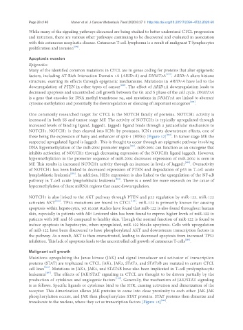
One commonly researched target for CTCL is the NOTCH family of proteins. NOTCH1 activity is
increased in both SS and tumor stage MF. The activity of NOTCH1 is typically upregulated through
increased levels of binding ligand, Jagged1. Jagged1 ligand binds through a juxtacellular mechanism to
NOTCH1. NOTCH1 is then cleaved into ICN1 by proteases. ICN1 exerts downstream effects, one of
these being the expression of hairy and enhancer of split-1 (HES1) [Figure 12] [202] . In tumor stage MF, the
suspected upregulated ligand is Jagged1. This is thought to occur through an epigenetic pathway involving
DNA hypermethylation of the miR-200c promoter region [202] . miR-200c can function as an oncogene that
inhibits activation of NOTCH1 through decreasing expression of the NOTCH1 ligand Jagged1. However,
hypermethylation in the promoter sequence of miR-200c decreases expression of miR-200c is seen in
MF. This results in increased NOTCH1 activity through an increase in levels of Jagged1 [202] . Overactivity
of NOTCH1 has been linked to decreased expression of PTEN and degradation of p53 in T-cell acute
lymphoblastic leukemia [203] . In addition, HES1 expression is also linked to the upregulation of the NF-κB
pathway in T-cell acute lymphoblastic leukemia [204] . There is a need for more research on the cause of
hypermethylation of these miRNA regions that cause downregulation.
NOTCH1 is also linked to the AKT pathway through PTEN and p53 regulation by miR-122. miR-122
activates AKT [205] . TP53 mutations are found in CTCL [199] . miR-122 is primarily known for causing
apoptosis within hepatocytes, yet recent studies have found that miR-122 is also found throughout human
skin, especially in patients with MF. Lesioned skin has been found to express higher levels of miR-122 in
patients with MF and SS compared to healthy skin. Though the normal function of miR-122 is found to
induce apoptosis in hepatocytes, when upregulated, miR-122 blocks apoptosis. Cells with upregulation
of miR-122 have been discovered to have phosphorylated AKT and downstream transcription factors in
the pathway. As a result, AKT is then overactivated, leading to decreased apoptosis from increased TP53
[205]
inhibition. This lack of apoptosis leads to the uncontrolled cell growth of cutaneous T-cells .
Malignant cell growth
Mutations upregulating the Janus kinase (JAK) and signal transducer and activator of transcription
proteins (STAT) are implicated in CTCL. JAK1, JAK3, STAT3, and STAT5B are mutated in certain CTCL
cell lines [206] . Mutations in JAK1, JAK3, and STAT5B have also been implicated in T-cell prolymphocytic
leukemia [207] . The effects of JAK/STAT signaling in CTCL are thought to be driven partially by the
production of cytokines and angiogenic factors [194] . Generally, the mechanism of JAK/STAT signaling
is as follows. Specific ligands or cytokines bind to the RTK, causing activation and dimerization of the
receptor. This dimerization allows JAK proteins to come into close proximity to each other. JAK-JAK
phosphorylation occurs, and JAK then phosphorylates STAT proteins. STAT proteins then dimerize and
translocate to the nucleus, where they act as transcription factors [Figure 12] [208] .