
Page 459 - Read Online
P. 459
Maner et al. J Cancer Metastasis Treat 2020;6:37 I http://dx.doi.org/10.20517/2394-4722.2020.60 Page 11 of 40
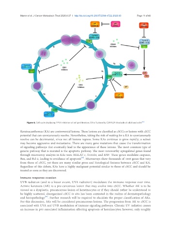
Figure 6. Cell cycle displaying TP53 inhibition of cell proliferation. G1 to S phase by CDKN2A blockade of cdk4 and cdk6 [81]
Keratoacanthomas (KA) are controversial lesions. These lesions are classified as cSCCs or lesions with cSCC
potential that can spontaneously resolve. Nevertheless, taking the risk of waiting for a KA to spontaneously
resolve can be detrimental, since not all lesions regress. Some KAs continue to grow rapidly; a subset
may become aggressive and metastasize. There are many gene mutations that cause the transformation
of signaling pathways that eventually lead to the appearance of these lesions. The most common type of
genetic pathway that is mutated is the apoptotic pathway. The most noteworthy upregulated genes found
through microarray analysis in KAs were MALAT-1, S100A8, and EHF. These genes modulate caspases,
[82]
Bax, and Bcl-2, leading to avoidance of apoptosis . Microarrays show thousands of 1449 genes that vary
from those of cSCC, yet there are many similar gross and histological features between cSCC and KA.
Regardless of this debate, KAs have a highly malignant potential similar to those of cSCC and should be
treated as soon as they are discovered.
Immune response evasion
UVB radiation (and to a lesser extent, UVA radiation) modulates the immune response over time.
Actinic keratosis (AK) is a pre-cancerous lesion that may evolve into cSCC. Whether AK is to be
viewed as a dysplastic, precancerous lesion of keratinocytes or if they should rather be understood to
be highly scattered, disorganized cSCC in situ has been contested in the realms of dermatopathology
[83]
and histopathology . Further research will be required to elucidate the proper classification of AKs.
For this discussion, AKs will be considered precancerous lesions. The progression from AK to cSCC is
associated with UVA and UVB modulation of immune signaling pathways. Chronic UV radiation causes
an increase in p53 associated inflammation affecting apoptosis of keratinocytes; however, only roughly