
Page 109 - Read Online
P. 109
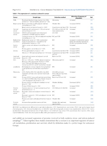
Page 4 of 12                        Schofield et al. J Cancer Metastasis Treat 2020;6:10  I  http://dx.doi.org/10.20517/2394-4722.2019.43

               Table 1. The expression of a-enolase is altered in cancer
                                                                                Alteration (frequency of
                Cancer                Sample type             Detection method                        Ref.
                                                                                     alteration)
                AML       Peripheral blood and bone marrow from AML   Microarray  Increased           [4]
                          patients (n = 41) and healthy patients (n = 20)
                Breast cancer Breast cancer tissue with adjacent matched   Western blot  Increased (100%)  [9]
                          normal tissue (n = 24)
                          Breast cancer and normal tissue (n = 244)  Quantitative real-time PCR Decreased  [10]
                Cervical   Squamous cell carcinoma (n = 33) and normal   2D-DIGE and MALDI-TOF  Increased  [40]
                cancer    cervical tissue (n = 17)         mass spectrometry
                Colorectal   SW620 cell line (lymph node metastasis)   2D-DIGE  Increased             [41]
                cancer    compared to SW480 (primary lesion)
                          Colorectal cancer (n = 48) and adjacent matched  IHC and PCR  Increased (56%)  [11]
                          normal tissue (n = 16)
                Endometrial  Endometrial cancer (n = 100), endometrial   IHC  Increased in endometrial cancer   [12]
                carcinoma  atypical hyperplasia (n = 22) and normal           (52%) and atypical hyperplasia
                          endometrium tissues (n = 20)                        (31.8%) compared to normal
                Gastric   Gastric cancer and adjacent normal tissue (n =   IHC  Increased             [13]
                cancer    94)
                          Primary gastric cancer tissue (n = 107)  IHC        Increased (48%)         [14]
                Glioma    Primary glioblastoma tissue (n = 24)  Quantitative real-time PCR Increased (68%)  [5]
                          Glioma (n = 136) and normal brain (n = 15) tissue Quantitative real-time PCR  Increased (69%)  [6]
                                                           and IHC
                Head and   Head and neck cancer and adjacent normal   Real-time PCR  Increased (50%)  [15]
                neck cancer  tissue (n = 44)
                HCC       HCC (n = 374 and n = 1309), adjacent matched   Microarray and IHC  Increased  [16]
                          tissue (n = 50), and normal tissue (n = 1442)
                          from TCGA and GEO data source; tissue
                          microarray (93 HCC and 87 normal liver tissues)
                Lymphoma  Pretherapeutic tumour biopsies from peripheral   IHC  Increased             [8]
                          T-cell lymphoma not otherwise classified (n =
                          87)
                Melanoma  A375, MeWo, MEL-HO, Colo-800, Colo-853   2D-DIGE and nano-  Increased       [7]
                          melanoma cell lines and a normal melanocyte   HPLC-chip ion trap mass
                          cell line                        spectrometry
                NSLC      Primary NSCLC tissue and matched normal lung  Real-time PCR and IHC  Increased  [17]
                          (n = 26) from RNA and primary NSCLC tissue (n
                          = 55) and normal lung tissue (n = 17)
                          Primary NSCLC and adjacent matched normal   Western blotting  Decreased (26%)  [42]
                          tissue (n = 46)
                          Primary NSCLS tissue (n = 36)    PCR                Increased (16%)         [18]
                Ovarian   Ovarian cancer (n = 4) and ovarian tissue from   2D-DIGE and LC-MS/MS  Increased  [19]
                cancer    endometriosis (n = 1)
                Pancreatic   Pancreatic cancer and adjacent normal tissue (n   IHC  Increased         [20]
                cancer    = 31)
                          Pancreatic cancer (n = 100) and adjacent normal  IHC  Increased (48%)       [21]
                          tissue (n = 80)
                          Primary pancreatic and adjacent normal tissue (n  Western blot  Increased   [22]
                          = 3)
                Prostate   Exosomes from prostate cancer cell lines  Western blot and mass   Decreased  [43]
                cancer                                     spectrometry

               2D-DIGE: two-dimensional differential in gel electrophoresis; HCC: hepatocellular carcinoma; HPLC: high performance liquid
               chromatography; IHC: immunohistochemistry; LC-MS/MS: liquid chromatography tandem mass spectrometry; MALDI-TOF: matrix
               assisted laser desorption/ionization time of flight; NSCLC: non-small cell lung cancer; PCR: polymerase chain reaction; AML: acute
               myeloid leukaemia; TCGA: the cancer genome atlas; GEO: gene expression omnibus


               and exhibit an increased expression of proteins involved in both oxidative stress- and sirtuin-induced
                        [62]
               autophagy . Taken together, these studies demonstrate that a-enolase is an important regulator of tumour
               cell metabolism, proliferation and survival, which by definition make it a perfect target for anticancer
               therapy.