Page 299 - Read Online
P. 299
Borniger. J Cancer Metastasis Treat 2019;5:23  I  http://dx.doi.org/10.20517/2394-4722.2018.107                             Page 3 of 18

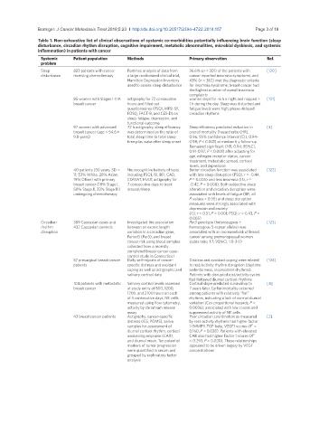
               Table 1. Non-exhaustive list of clinical observations of systemic co-morbidities potentially influencing brain function (sleep
               disturbance, circadian rhythm disruption, cognitive impairment, metabolic abnormalities, microbial dysbiosis, and systemic
               inflammation) in patients with cancer
                Systemic    Patient population   Methods                Primary observation          Ref.
                problem
                Sleep       823 patients with cancer   Post-hoc analysis of data from   36.6% (n = 301) of the patients with   [120]
                disturbance  receiving chemotherapy  a large randomized clinical trial;   cancer reported insomnia symptoms, and
                                                 Hamilton Depression Inventory   43% (n = 362) met the diagnostic criteria
                                                 used to assess sleep disturbance   for insomnia syndrome; breast cancer had
                                                                        the highest number of overall insomnia
                                                                        complaints
                            85 women with Stages I-IIIA   actigraphy for 72 consecutive   women slept for ~6 h a night and napped >   [121]
                            breast cancer        hours and filled out   1 h during the day. Sleep was disturbed and
                                                 questionnaires (PSQI, MFSI-SF,   fatigue levels were high; phase-delayed
                                                 FOSQ, FACT-B, and CES-D) on   circadian rhythms
                                                 sleep, fatigue, depression, and
                                                 functional outcome
                            97 women with advanced   72 h actigraphy; sleep efficiency   Sleep efficiency predicted reduction in   [3]
                            breast cancer (age = 54.6 ±   was determined as the ratio of   overall mortality [hazard ratio (HR),
                            9.8 years)           total sleep time to total sleep   0.96; 95% confidence interval (CI), 0.94-
                                                 time plus wake after sleep onset  0.98; P < 0.001] at median 6 y follow-up.
                                                                        Remained significant (HR, 0.94; 95%CI,
                                                                        0.91-0.97; P < 0.001) after adjusting for
                                                                        age, estrogen receptor status, cancer
                                                                        treatment, metastatic spread, cortisol
                                                                        levels, and depression
                            40 patients (50 years, SD =   Neurocognitive battery of tests   Better circadian function was associated   [122]
                            11; 53% White, 28% Asian,   including PSQI, ISI, BFI, CAD,   with less sleep disruption (PSQI, r = -0.44,
                            19% Other) with primary   COWAT, HVLT; actigraphy for   P = 0.005) and less insomnia (ISI, r =
                            breast cancer (18% Stage I,   7 consecutive days to track   -0.42, P = 0.008). Both subjective sleep
                            50% Stage II, 33% Stage III)   arousal/sleep  alteration and circadian disruption were
                            undergoing chemotherapy                     associated with levels of fatigue (BFI, all
                                                                        P-values < 0.05) and sleep disruption
                                                                        measures were strongly associated with
                                                                        depression and anxiety
                                                                        (ISI: r = 0.51, P = 0.001; PSQI: r = 0.43, P =
                                                                        0.005)
                Circadian   389 Caucasian cases and   Investigated the association   Per3 genotype (heterozygous +   [123]
                rhythm      432 Caucasian controls  between an exonic length   homozygous 5-repeat alleles) was
                disruption                       variation in a circadian gene,   associated with an increased risk of breast
                                                 Period3 (Per3), and breast   cancer among premenopausal women
                                                 cancer risk using blood samples   (odds ratio, 1.7; 95%CI, 1.0-3.0)
                                                 collected from a recently
                                                 completed breast cancer case-
                                                 control study in Connecticut
                            57 presurgical breast cancer   Daily self-reports of cancer-  Distress and avoidant coping were related   [124]
                            patients             specific distress and avoidant   to rest/activity rhythm disruption (daytime
                                                 coping as well as actigraphic and   sedentariness, inconsistent rhythms).
                                                 salivary cortisol data  Patients with disrupted rest/activity cycles
                                                                        had flattened diurnal cortisol rhythms
                            104 patients with metastatic   Salivary cortisol levels assessed   Cortisol slope predicted survival up to   [4]
                            breast cancer        at study entry at 800, 1200,   7 years later. Earlier mortality occurred
                                                 1700, and 2100 hours on each   among patients with relatively “flat”
                                                 of 3 consecutive days; NK cells   rhythms, indicating a lack of normal diurnal
                                                 measured using flow cytometry,   variation (Cox proportional hazards, P =
                                                 activity by chromium release   0.0036); associated with low counts and
                                                 assay                  suppressed activity of NK cells
                            43 breast cancer patients  Actigraphy, cancer-specific   Poor circadian coordination as measured   [2]
                                                 distress (IES, POMS), saliva   by rest-activity rhythms had higher factor
                                                                                               2
                                                 samples for assessment of   1 (MMP9, TGF-beta, VEGF) scores (R  =
                                                 diurnal cortisol rhythm, cortisol   0.160, P = 0.038). Patients with elevated
                                                 awakening response (CAR),   CAR also had higher Factor 1 scores (R   2
                                                 and diurnal mean. Ten potential   = 0.293, P = 0.020). These relationships
                                                 markers of tumor progression   appeared to be driven largely by VEGF
                                                 were quantified in serum and   concentrations
                                                 grouped by exploratory factor
                                                 analysis

