Page 303 - Read Online
P. 303
Borniger. J Cancer Metastasis Treat 2019;5:23  I  http://dx.doi.org/10.20517/2394-4722.2018.107                             Page 7 of 18


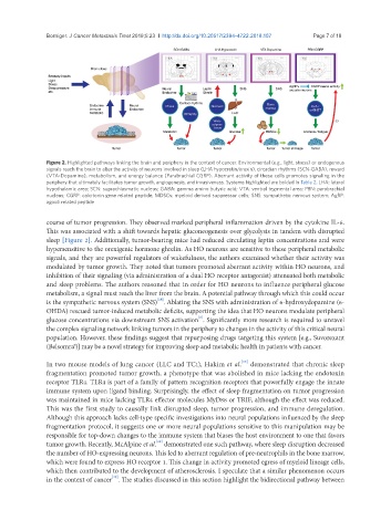
               Figure 2. Highlighted pathways linking the brain and periphery in the context of cancer. Environmental (e.g., light, stress) or endogenous
               signals reach the brain to alter the activity of neurons involved in sleep (LHA hypocretin/orexin), circadian rhythms (SCN-GABA), reward
               (VTA-Dopamine), metabolism, and energy balance (Parabrachial CGRP). Aberrant activity of these cells promotes signaling in the
               periphery that ultimately facilitates tumor growth, angiogenesis, and invasiveness. Systems highlighted are bolded in Table 2. LHA: lateral
               hypothalamic area; SCN: suprachiasmatic nucleus; GABA: gamma amino butyric acid; VTA: ventral tegmental area; PBN: parabrachial
               nucleus; CGRP: calcitonin gene related peptide; MDSCs: myeloid derived suppressor cells; SNS: sympathetic nervous system; AgRP:
               agouti related peptide

               course of tumor progression. They observed marked peripheral inflammation driven by the cytokine IL-6.
               This was associated with a shift towards hepatic gluconeogenesis over glycolysis in tandem with disrupted
               sleep [Figure 2]. Additionally, tumor-bearing mice had reduced circulating leptin concentrations and were
               hypersensitive to the orexigenic hormone ghrelin. As HO neurons are sensitive to these peripheral metabolic
               signals, and they are powerful regulators of wakefulness, the authors examined whether their activity was
               modulated by tumor growth. They noted that tumors promoted aberrant activity within HO neurons, and
               inhibition of their signaling (via administration of a dual HO receptor antagonist) attenuated both metabolic
               and sleep problems. The authors reasoned that in order for HO neurons to influence peripheral glucose
               metabolism, a signal must reach the liver from the brain. A potential pathway through which this could occur
                                                 [35]
               is the sympathetic nervous system (SNS) . Ablating the SNS with administration of 6-hydroxydopamine (6-
               OHDA) rescued tumor-induced metabolic deficits, supporting the idea that HO neurons modulate peripheral
                                                             [6]
               glucose concentrations via downstream SNS activation . Significantly more research is required to unravel
               the complex signaling network linking tumors in the periphery to changes in the activity of this critical neural
               population. However, these findings suggest that repurposing drugs targeting this system [e.g., Suvorexant
               (Belsomra®)] may be a novel strategy for improving sleep and metabolic health in patients with cancer.

                                                                          [13]
               In two mouse models of lung cancer (LLC and TC1), Hakim et al.  demonstrated that chronic sleep
               fragmentation promoted tumor growth, a phenotype that was abolished in mice lacking the endotoxin
               receptor TLR4. TLR4 is part of a family of pattern recognition receptors that powerfully engage the innate
               immune system upon ligand binding. Surprisingly, the effect of sleep fragmentation on tumor progression
               was maintained in mice lacking TLR4 effector molecules MyD88 or TRIF, although the effect was reduced.
               This was the first study to causally link disrupted sleep, tumor progression, and immune deregulation.
               Although this approach lacks cell-type specific investigations into neural populations influenced by the sleep
               fragmentation protocol, it suggests one or more neural populations sensitive to this manipulation may be
               responsible for top-down changes to the immune system that biases the host environment to one that favors
                                                 [49]
               tumor growth. Recently, McAlpine et al.  demonstrated one such pathway, where sleep disruption decreased
               the number of HO-expressing neurons. This led to aberrant regulation of pre-neutrophils in the bone marrow,
               which were found to express HO receptor 1. This change in activity promoted egress of myeloid lineage cells,
               which then contributed to the development of atherosclerosis. I speculate that a similar phenomenon occurs
                                   [49]
               in the context of cancer . The studies discussed in this section highlight the bidirectional pathway between

