
Page 584 - Read Online
P. 584
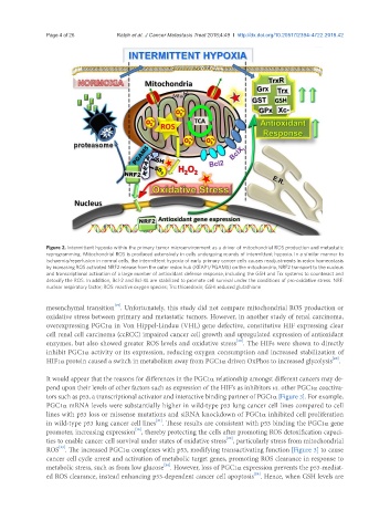
Page 4 of 26                              Ralph et al. J Cancer Metastasis Treat 2018;4:49  I  http://dx.doi.org/10.20517/2394-4722.2018.42


               Figure 2. Intermittent hypoxia within the primary tumor microenvironment as a driver of mitochondrial ROS production and metastatic
               reprogramming. Mitochondrial ROS is produced extensively in cells undergoing rounds of intermittent hypoxia. In a similar manner to
               ischaemia/reperfusion in normal cells, the intermittent hypoxia of early primary cancer cells causes readjustments in redox homeostasis
               by increasing ROS activated NRF2 release from the outer redox hub (KEAP1/PGAM5) on the mitochondria, NRF2 transport to the nucleus
               and transcriptional activation of a large number of antioxidant defense response, including the GSH and Trx systems to counteract and
               detoxify the ROS. In addition, Bcl-2 and Bcl-XL are stabilized to promote cell survival under the conditions of pro-oxidative stress. NRF:
               nuclear respiratory factor; ROS: reactive oxygen species; Trx: thioredoxin; GSH: reduced glutathione

                                    [19]
               mesenchymal transition . Unfortunately, this study did not compare mitochondrial ROS production or
               oxidative stress between primary and metastatic tumors. However, in another study of renal carcinoma,
               overexpressing PGC1α in Von Hippel-Lindau (VHL) gene defective, constitutive HIF expressing clear
               cell renal cell carcinoma (ccRCC) impaired cancer cell growth and upregulated expression of antioxidant
                                                                         [20]
               enzymes, but also showed greater ROS levels and oxidative stress . The HIFs were shown to directly
               inhibit PGC1α activity or its expression, reducing oxygen consumption and increased stabilization of
                                                                                                      [20]
               HIF1α protein caused a switch in metabolism away from PGC1α driven OxPhos to increased glycolysis .
               It would appear that the reasons for differences in the PGC1α relationship amongst different cancers may de-
               pend upon their levels of other factors such as expression of the HIF’s as inhibitors vs. other PGC1α coactiva-
               tors such as p53, a transcriptional activator and interactive binding partner of PGC1α [Figure 3]. For example,
               PGC1α mRNA levels were substantially higher in wild-type p53 lung cancer cell lines compared to cell
               lines with p53 loss or missense mutations and siRNA knockdown of PGC1α inhibited cell proliferation
                                                 [21]
               in wild-type p53 lung cancer cell lines . These results are consistent with p53 binding the PGC1α gene
                                          [16]
               promoter, increasing expression , thereby protecting the cells after promoting ROS detoxification capaci-
                                                                      [22]
               ties to enable cancer cell survival under states of oxidative stress , particularly stress from mitochondrial
                   [23]
               ROS . The increased PGC1α complexes with p53, modifying transactivating function [Figure 3] to cause
               cancer cell cycle arrest and activation of metabolic target genes, promoting ROS clearance in response to
                                                   [24]
               metabolic stress, such as from low glucose . However, loss of PGC1α expression prevents the p53-mediat-
                                                                               [24]
               ed ROS clearance, instead enhancing p53-dependent cancer cell apoptosis . Hence, when GSH levels are