
Page 201 - Read Online
P. 201
Galletti et al. Using CTCs in prostate cancer
Taking advantage of the larger size of CTCs compared those undergone EMT potentially missed by epithelial
to hematopoietic cells (15-25 µm vs. less than 12 µm), antigen-based approaches. In a direct comparison
many different microfiltration devices have been of performance in CTC enumeration in breast, lung
developed and tested clinically for the isolation of and prostate cancers, the ISET assay isolated CTCs
CTCs. These devices employ small pore membranous in higher numbers than CellSearch , suggesting that
®
filters that select CTCs apart from the contaminating size-based methods could isolate more than the
PBMCs by size . ScreenCell has developed a merely EpCAM positive CTCs . The ability of ISET
®
[21]
[24]
range of devices based on microporous membrane to retain CTCs with EMT molecular features is more
filters, which are engineered to either capture CTCs directly supported by other evidence in the literature
for cytological studies, molecular and genetic analysis, that show how ISET-isolated CTCs can express
or for CTC culture in vitro . Another largely clinically antigens of mesenchymal origins with concomitant
[22]
used filter-based approach, ISET (Isolation by Size of lack of epithelial markers [25,26] . In addition, all these
®
Epithelial Tumor cells, Rare cells Diagnostics), uses size-based approaches provide the advantage of
membranes with 8 µm pores to retain CTCs allowing isolating CTC-clusters, which proved to be critical in
smaller blood cells to pass through and be discarded . metastasis initiation .
[23]
[27]
Overall, all filtration-based CTC isolation techniques Similarly to simple density gradient centrifugation,
have the advantage of being “antigen agnostic”; microfiltration devices produce live, unaltered CTCs
as these methods do not discriminate based on with the added benefit of higher purity. These CTCs
expression of plasma membrane antigens, molecularly lend themselves to a wide variety of downstream
diverse CTC subpopulations can be retained, including assays, which can reveal clinically meaningful
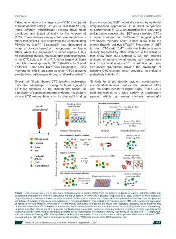
Figure 1: Descriptive overview of the main methodologies to isolate CTCs from the peripheral blood of cancer patients. CTCs can
be isolated and enriched from the contaminating WBCs based on either their physical properties (e.g. size, density) or their biological
properties (i.e. expression of tumor-selective markers on the plasma membrane). Physical property-based techniques have the potential
advantage of isolating molecularly heterogeneous CTC subpopulations, thus including CTCs undergone EMT with low/absent expression
of epithelial surface markers. Presence of contaminating leukocytes represents the major limit. Biological property-based methods rely
on positive selection of CTCs based on the expression of cancer-specific markers on the surface of circulating tumor cells; alternatively,
negative depletion leaves out the unwanted contaminating leukocytes, based on immunomediated depletion of cells expressing the
leukocyte-specific CD45 marker. Biological property-based technologies are characterized by high purity of the obtained CTC population,
with the caveat of missing CTC subpopulations lacking the expression of the surface marker when positive selection is adopted. CTC:
circulating tumor cell; EMT: epithelial-mesenchymal transition; WBC: white blood cells; RBC: red blood cell
Journal of Cancer Metastasis and Treatment ¦ Volume 3 ¦ September 27, 2017 193