
Page 403 - Read Online
P. 403
Shah et al. Breast metastasis mimicking as second primary cancer
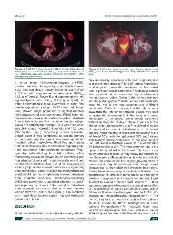
Figure 4: FDG-PET scan showed FDG avid soft tissue density Figure 5: FDG avid hypermetabolic right inguinal lymph node
lesion (size 4.2 cm × 2.8 cm SUV max 13.2) in left breast. FDG- SUV max - 5.1. FDG: fluorodeoxyglucose; SUV: standardized uptake
PET: fluorodeoxyglucose-positron emission tomography; SUV: value
standardized uptake value
they are usually associated with poor prognosis, due
a whole body 18-fluorodeoxyglucose (18-FDG) to disseminated disease. It is of utmost importance
[7]
positron emission tomography scan which showed to distinguish metastatic carcinoma to the breast
FDG avid soft tissue density lesion of size 4.2 cm from a primary breast carcinoma. Metastatic spread
[8]
× 2.8 cm with standardized uptake value (SUV) max from anorectal cancer occurs both by lymphatic and
13.2 in left breast [Figure 4] and hypermetabolic right hematogenous routes. Owing to the venous drainage
inguinal lymph node SUV max 5.1 [Figure 5] with no into the portal system from the superior hemorrhoidal
other hypermetabolic focus elsewhere in body. Fine vein, the liver is the most common site of distant
needle aspiration cytology (FNAC) from left breast metastasis. Systemic drainage into the inferior vena
lump showed single population of atypical epithelial cava from the inferior hemorroidal plexus may lead
cells suggestive of adenocarcinoma. FNAC from right to metastatic involvement of the lung and bone.
inguinal node was also done which reported metastasis Metastases to the breast from anorectal carcinoma
from adenocarcinoma. Her carcinoembryonic antigen without involvement of any of these organs is a rare
(CEA) and carbohydrate antigen-15.3 was done which phenomenon. Schaekelford et al. reviewed 19 cases
[8]
was 26.8 ng/mL (Normal 0-4 ng/mL) and 17.2 u/mL of colorectal carcinoma metastasizing to the breast
(Normal 0-35 u/mL) respectively. In view of isolated and reported a majority of cases with metastases to the
breast lesion it was considered as second primary left breast 55%, with the right breast 30% and 3 cases
of the breast and the patient was taken up for left with bilateral breast metastasis. In our case, patient
modified radical mastectomy. Right iliac and inguinal had left breast metastasis similar to the observation
node dissection was also performed for regional lymph by Schaekelford et al. The most common site is the
[9]
node recurrence from carcinoma anorectum. Post- upper outer quadrant of the breast. They can occur
operative histopathology from left modified radical as synchronous lesions or may follow the primary by
mastectomy specimen showed mucin secreting signet months to years. Metastatic breast lesions are typically
ring adenocarcinoma with lymphovascular emboli and mobile, well demarcated, firm, rapidly growing, discrete
lymphocytic infiltration. Nine out of 16 dissected left masses and may be confused with benign breast
axillary lymph nodes showed metastasis of signet ring disease due to their often well-circumscribed nature.
adenocarcinoma. Six out of 8 right inguinal lymph nodes Rarely these lesions may be multiple or bilateral. The
and 2 out of 4 right iliac lymph nodes showed metastasis interpretation is difficult in some cases so a history of
from anorectal carcinoma. Immunohistochemistry previous malignancy is important for the radiologist
(IHC) was performed to ascertain whether the lesion in order to evaluate these breast lesions. [10,11] Other
was a primary carcinoma of the breast or metastasis features suggestive of metastasis to breast are location
from anorectal carcinoma. Result of IHC markers of the lump in either fat or subcutaneous tissue, lack of
was as shown in Table 1 and Figure 6. IHC combined micro-calcification in mammogram and lack of in situ
with morphology favored signet ring cell metastatic disease on histopathological examination. [12,13] The
carcinoma to breast. correct diagnosis is therefore crucial in these patients
so as to decide the further management of these
DISCUSSION patients. Histopathology for metastatic lesion may
be invasive adenocarcinoma, often with mucinous or
Breast metastases from colon cancer are very rare and signet-ring cell features, but unlike primary lesion of the
Journal of Cancer Metastasis and Treatment ¦ Volume 2 ¦ September 30, 2016 393