
Page 402 - Read Online
P. 402
Shah et al. Breast metastasis mimicking as second primary cancer
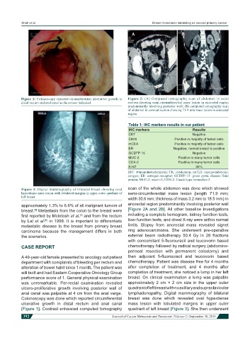
Figure 1: Colonoscopy reported circumferential ulcerative growth in Figure 2: (A) Computed tomography scan of abdomen in axial
distal rectum and anal canal as the arrows indicated section showing semi-circumferential mass lesion in anorectal region
predominantly involving posterior wall; (B) computed tomography scan
of abdomen in coronal section showing 71.9 mm mass lesion in anorectal
region
Table 1: IHC markers results in our patient
IHC markers Results
CK7 Negative
CK20 Positive in majority of tumor cells
mCEA Positive in majority of tumor cells
ER Negative, normal breast is positive
GCDFP-15 Negative
MUC-2 Positive in many tumor cells
CDX-2 Positive in many tumor cells
Ki-67 30%
IHC: immunohistochemistry; CK: cytokeratin; mCEA: carcinoembryonic
antigen; ER: estrogen receptor; GCDFP-15: gross cystic disease fluid
protein; MUC-2: mucin-2; CDX-2: Caudal type homeobox-2
Figure 3: Digital mammography of bilateral breast showing oval scan of the whole abdomen was done which showed
hyperdense mass lesion with lobulated margins in upper outer quadrant of semi-circumferential mass lesion (length 71.9 mm;
left breast
width 30.6 mm; thickness of mass 3.2 mm to 18.5 mm) in
approximately 1.3% to 6.6% of all malignant tumors of anorectal region predominantly involving posterior wall
breast. Metastasis from the colon to the breast were [Figure 2A and 2B]. All other baseline investigations
[4]
first reported by McIntosh et al. and from the rectum including a complete hemogram, kidney function tests,
[5]
by Lal et al. in 1999. It is important to differentiate liver function tests, and chest X-ray were within normal
[6]
metastatic disease to the breast from primary breast limits. Biopsy from anorectal mass revealed signet
carcinoma because the management differs in both ring adenocarcinoma. She underwent pre-operative
the scenarios. external beam radiotherapy 50.4 Gy in 28 fractions
with concomitant 5-fluorouracil and leucovorin based
CASE REPORT chemotherapy followed by radical surgery (abdomino-
perineal resection with permanent colostomy) and
A 49-year-old female presented to oncology out patient then adjuvant 5-fluorouracil and leucovorin based
department with complaints of bleeding per rectum and chemotherapy. Patient was disease free for 4 months
alteration of bowel habit since 1 month. The patient was after completion of treatment, and 4 months after
will built and had Eastern Cooperative Oncology Group completion of treatment, she noticed a lump in her left
performance score of 1. General physical examination breast. On clinical examination a lump was palpable
was unremarkable. Per-rectal examination revealed approximately 2 cm × 2 cm size in the upper outer
ulcero-proliferative growth involving posterior wall of quadrant of left breast with no axillary and supraclavicular
anal canal was palpable at 4 cm from the anal verge. lymphadenopathy. Digital mammography of bilateral
Colonoscopy was done which reported circumferential breast was done which revealed oval hyperdense
ulcerative growth in distal rectum and anal canal mass lesion with lobulated margins in upper outer
[Figure 1]. Contrast enhanced computed tomography quadrant of left breast [Figure 3]. She then underwent
392 Journal of Cancer Metastasis and Treatment ¦ Volume 2 ¦ September 30, 2016