
Page 387 - Read Online
P. 387
Adachi et al. Pediatric gliomatosis cerebri
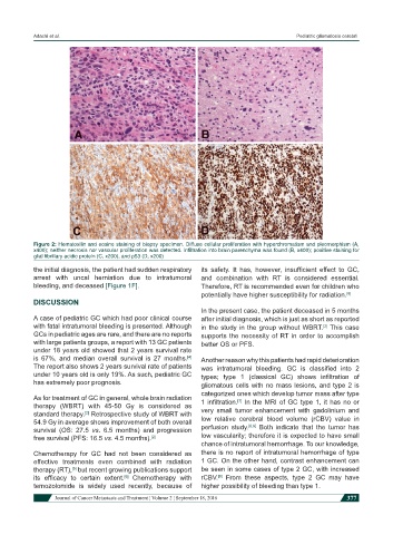
Figure 2: Hematoxilin and eosine staining of biopsy specimen. Diffuse cellular proliferation with hyperchromatism and pleomorphism (A,
x400); neither necrosis nor vascular proliferation was detected. Infiltration into brain parenchyma was found (B, x400); positive staining for
glial fibrillary acidic protein (C, x200), and p53 (D, x200)
the initial diagnosis, the patient had sudden respiratory its safety. It has, however, insufficient effect to GC,
arrest with uncal herniation due to intratumoral and combination with RT is considered essential.
bleeding, and deceased [Figure 1F]. Therefore, RT is recommended even for children who
potentially have higher susceptibility for radiation. [6]
DISCUSSION
In the present case, the patient deceased in 5 months
A case of pediatric GC which had poor clinical course after initial diagnosis, which is just as short as reported
with fatal intratumoral bleeding is presented. Although in the study in the group without WBRT. This case
[2]
GCs in pediatric ages are rare, and there are no reports supports the necessity of RT in order to accomplish
with large patients groups, a report with 13 GC patients better OS or PFS.
under 18 years old showed that 2 years survival rate
is 67%, and median overall survival is 27 months. Another reason why this patients had rapid deterioration
[4]
The report also shows 2 years survival rate of patients was intratumoral bleeding. GC is classified into 2
under 10 years old is only 19%. As such, pediatric GC types; type 1 (classical GC) shows infiltration of
has extremely poor prognosis. gliomatous cells with no mass lesions, and type 2 is
categorized ones which develop tumor mass after type
As for treatment of GC in general, whole brain radiation 1 infiltration. In the MRI of GC type 1, it has no or
[7]
therapy (WBRT) with 45-50 Gy is considered as
standard therapy. Retrospective study of WBRT with very small tumor enhancement with gadolinium and
[2]
54.9 Gy in average shows improvement of both overall low relative cerebral blood volume (rCBV) value in
[8,9]
survival (OS: 27.5 vs. 6.5 months) and progression perfusion study. Both indicate that the tumor has
free survival (PFS: 16.5 vs. 4.5 months). [2] low vascularity; therefore it is expected to have small
chance of intratumoral hemorrhage. To our knowledge,
Chemotherapy for GC had not been considered as there is no report of intratumoral hemorrhage of type
effective treatments even combined with radiation 1 GC. On the other hand, contrast enhancement can
therapy (RT), but recent growing publications support be seen in some cases of type 2 GC, with increased
[5]
its efficacy to certain extent. Chemotherapy with rCBV. From these aspects, type 2 GC may have
[9]
[6]
temozolomide is widely used recently, because of higher possibility of bleeding than type 1.
Journal of Cancer Metastasis and Treatment ¦ Volume 2 ¦ September 18, 2016 377