
Page 386 - Read Online
P. 386
Adachi et al. Pediatric gliomatosis cerebri
cerebellum, and spinal cord, affecting both gray and detected. Tumor cell infiltration in the peripheral zone of
white matter. It is classified as grade IV in World Health a tumor was found [Figure 2B]. Immunohistochemistry
Organization 2007 criteria, regardless of its histo- revealed positive staining for glial fibrillary acidic
pathological features. protein, and nuclear staining of p53. MIB-1 proliferation
index was about 50% [Figure 2C and 2D]. With these
GCs in most cases are seen in the adult population, results, histopathological diagnosis was made as
rarely suffering young age group. There are two peaks anaplastic astrocytoma (grade III). The final clinical
of patients’ age distribution in the second decades and diagnosis was determined as gliomatosis cerebri due
forties. [3] to invasion into 3 cerebral lobes and brainstem.
We report here a pediatric patient with GC who Considering potential poor prognosis of the disease,
refused any treatment, subsequently followed by rapid the patient’s parents refused either radiation or
deterioration with intratumoral bleeding. chemotherapy, and only oral corticosteroid and
rehabilitation was given to the patient. Five months after
CASE REPORT
A 14-year-old woman presented with generalized
tonic-clonic seizures following to history of morning
headache, and mild cognitive deteriorations. She
had noticed slowly progressing weakness on her
right face and upper extremity, and numbness on
her right side.
Neurological examination on admission revealed
right facial droop and pronator drift on the right
side. The patient originally was right-handed active
softball player, but grasping power was weak with
21 kg on the right and 29 kg on the left at the time.
Decreased proprioception and touch sensation was
observed both in upper and lower extremities on
the right. She was previously healthy and achieved
normal developmental milestones and scholastic
achievement up to an onset, but had experienced
a decline in cognition. Wechsler Intelligence Scale
for Children (WISC)-IV score shows intelligence
quotient (IQ) 50.
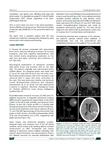
Fluid-attenuated inversion recovery (FLAIR) and T2-
wighted sequences of magnetic resonance imaging
(MRI) showed hyperintense signal lesion in the white
matter of the left frontal, parietal, and temporal lobes
[Figure 1A and 1B]. Follow up MRI in 2 months later
showed development of the lesion into the frontal lobe,
as well as the brainstem and corpus callosum [Figure
1C and 1D]. MR spectroscopy at the time shows high
peaks in both chorine (Cho)/N-acetilaspartate (NAA)
(2.9) and Cho/Creatine (Cr) (2.46), suggesting high
grade glioma [Figure 1E].
Open biopsy was performed targeting occipito-
temporal mass near posterior horn of the left lateral Figure 1: FLAIR and T2 weighted (T2WI) MR images demonstrating
hyperintense area into 3 cerebral lobes, corpus callosum, and
ventricle, which shows solid swelling without contrast brainstem (A: axial FLAIR; B: coronal T2WI); follow-up FLAIR
enhancement on MRI. Hematoxilin and eosine MRI took 2 months after initial symptoms (C: axial; D: coronal);
staining of specimen shows marked cellularity, with MR spectroscopy suggesting high-grade glioma (E); axial CT
scan demonstrating intratumoral hemorrhage 5 months after initial
marked hyperchromatism and pleomorphism [Figure symptoms (F). FLAIR: fluid-attenuated inversion recovery; MRI:
2A]. Neither necrosis nor vascular proliferation was magnetic resonance imaging; CT: computed tomography
376 Journal of Cancer Metastasis and Treatment ¦ Volume 2 ¦ September 18, 2016