
Page 235 - Read Online
P. 235
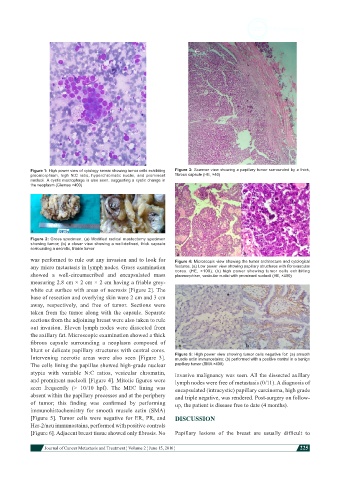
Figure 1: High power view of cytology smear showing tumor cells exhibiting   Figure 3: Scanner view showing a papillary tumor surrounded by a thick,
            pleomorphism, high N:C ratio, hyperchromatic nuclei, and prominent   fibrous capsule (HE, ×40)
            nucleoli. A cystic macrophage is also seen, suggesting a cystic change in
            the neoplasm (Giemsa ×400)


            Figure 2: Gross specimen. (a) Modified radical mastectomy specimen
            showing tumor; (b) a closer view showing a well-defined, thick capsule
            sorrounding a necrotic, friable tumor
            was performed to rule out any invasion and to look for   Figure 4: Microscopic view showing the tumor architecture and cytological
            any micro metastasis in lymph nodes. Gross examination   features. (a) Low power view showing papillary structures with fibrovascular
            showed a well-circumscribed and encapsulated mass   cores.  (HE,  ×100);  (b)  high  power  showing  tumor  cells  exhibiting
                                                               pleomorphism, vesicular nuclei with prominent nucleoli (HE, ×400)
            measuring 2.8 cm × 2 cm × 2 cm having a friable grey-
            white cut surface with areas of necrosis [Figure 2]. The
            base of resection and overlying skin were 2 cm and 3 cm
            away, respectively, and free of tumor. Sections were
            taken from the tumor along with the capsule. Separate
            sections from the adjoining breast were also taken to rule
            out invasion. Eleven lymph nodes were dissected from
            the axillary fat. Microscopic examination showed a thick
            fibrous  capsule  surrounding  a  neoplasm  composed  of
            blunt or delicate papillary structures with central cores.
            Intervening necrotic areas were also seen [Figure 3].   Figure 5: High power view showing tumor cells negative for: (a) smooth
                                                               muscle actin immunostains; (b) performed with a positive control in a benign
            The cells lining the papillae showed high-grade nuclear   papillary tumor (SMA ×400)
            atypia with variable N:C ratios, vesicular chromatin,   invasive malignancy was seen. All the dissected axillary
            and prominent nucleoli [Figure 4]. Mitotic figures were   lymph nodes were free of metastasis (0/11). A diagnosis of
            seen  frequently  (>  10/10  hpf).  The  MEC  lining  was   encapsulated (intracystic) papillary carcinoma, high grade
            absent within the papillary processes and at the periphery   and triple negative, was rendered. Post-surgery on follow-
            of  tumor;  this  finding  was  confirmed  by  performing   up, the patient is disease free to date (4 months).
            immunohistochemistry  for  smooth  muscle  actin  (SMA)
            [Figure 5].  Tumor cells were negative for ER, PR, and   DISCUSSION
            Her-2/neu immunostains, performed with positive controls
            [Figure 6]. Adjacent breast tissue showed only fibrosis. No   Papillary  lesions  of  the  breast  are  usually  difficult  to


                        Journal of Cancer Metastasis and Treatment ¦ Volume 2 ¦ June 15, 2016 ¦           225