Page 231 - Read Online
P. 231
to  periodontal/dental  infection  and  disease,  will  lead  to
            exposure of the alveolar bone, which may already be necrotic
            and will not heal.  Infection, within a unifying concept
                           [17]
            of  medication-related  impaired  immune  response  was
            proposed to play an important role in the pathophysiology
            of ONJ. [18]


            In this paper we report what we believe is the first case of
            ONJ in a patient with acute myeloid leukemia (AML), who
            was treated with azacitidine. The presence of alveolar bone
            disease leads to the dental extraction and the subsequent
            diagnosis of ONJ.
            CASE REPORT
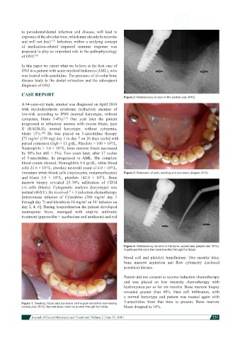
                                                               Figure 2: Radiolucency is seen in the socket (July 2015)
            A 64-year-old male, smoker was diagnosed on April 2010
            with  myelodysplastic  syndrome  (refractory  anemia)  of
            low-risk  according  to  IPSS  (normal  karyotype,  without
            cytopenia,  blasts  3-4%).  One year later the patient
                                 [19]
            progressed  to  refractory  anemia  with  excess  blasts,  type
            II  (RAEB-II),  normal  karyotype,  without  cytopenia,
            blasts 15%.   He  was  placed  on  5-azacitidine  therapy
                      [20]
            [75 mg/m  (150 mg) day 1 to day 7 on 28 days cycle] with
                    2
            partial remission (Hgb > 11 g/dL, Platelets > 100 × 10 /L,
                                                        9
            Neutrophils > 1.0 × 10 /L, bone marrow blasts decreased
                               9
            by 50% but still > 5%). Two years later, after 17 cycles
            of  5-azacitidine,  he  progressed  to  AML.  His  complete
            blood counts showed: Hemoglobin 9.6 gr/dL, white blood
            cells 21.6 × 10 /L, absolute neutrofil count of 4.0 × 10 /L,
                        9
                                                        9
            immature white blood cells (myelocytes, metamyelocytes)   Figure 3: Remission of pain, swelling and purulence (August 2015)
            and blasts 5.0  ×  10 /L,  platelets  142.0  ×  10 /L.  Bone
                             9
                                                   9
            marrow  biopsy  revealed  25-30%  infiltration  of  CD34
            (+)  cells  (blasts).  Cytogenetic  analysis  (karyotype)  was
            normal (46XY). He received 7 + 3 induction chemotherapy
            [intravenous  infusion  of  Cytarabine  (200  mg/m  day  1
                                                     2
            through day 7) and Idarubicin 10 mg/m  on 30’ infusion on
                                           2
            day 2, 4, 6]. During hospitalization the patient developed
            neutropenic  fever,  managed  with  empiric  antibiotic
            treatment (piperacilin + tazobactam and amikacin) and red


                                                               Figure 4: Radiolucency remains in the bone, socket area (September 2015).
                                                               A gutta-percha cone has been inserted through the fistula
                                                               blood  cell  and  platelets  transfusions.  Two  months  later,
                                                               bone  marrow  aspiration  and  flow  cytometry  disclosed
                                                               persistent disease.

                                                               Patient did not consent to receive induction chemotherapy
                                                               and  was  placed  on  low  intensity  chemotherapy  with
                                                               hydroxyurea per os for six months. Bone marrow biopsy
                                                               revealed  greater  than  60%  blast  cell  infiltration,  with
                                                               a  normal  karyotype  and  patient  was  treated  again  with
                                                               5-azacitidine  from  that  time  to  present.  Bone  marrow
            Figure 1: Swelling, fistula and purulunce on the post extraction non-healing
            socket (July 2015). Necrotic bone could be probed through the fistula  blasts dropped to 14%.


                        Journal of Cancer Metastasis and Treatment ¦ Volume 2 ¦ June 15, 2016 ¦           221

