
Page 239 - Read Online
P. 239
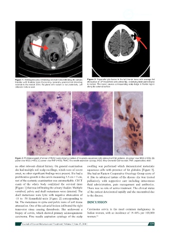
Figure 1: Heterogeneously enhancing cervical mass infiltrating the urinary   Figure 2: Expansile lytic lesion in the left frontal bone with average fat
            bladder with bladder base thickening. Laterally parametrial stranding   attenuation of -27 Hounsfield units (arrow tip). Underlying brain parenchyma
            extends to the medial 2/3rd. Fat plane with rectum is lost posteriorly. Left   is normal. The lesion causes corresponding scalp bulge in frontal region
            obturator node is seen                             along the external surface


            Figure 3: Photomicrograph of smear of FNAC scalp showing clusters of neoplastic squamous cells admixed with fat globules. (A) power view MGG (×100); (B)
            power view MGG (×400); (C) power view PAP (×100). FNAC: fine needle aspiration cytology; MGG: May-Grunwald Geimsa stain; PAP: papanicolaou stain
            no other relevant clinical history. On general examination   swelling was performed which demonstrated metastatic
            she had multiple soft scalp swellings, which were of recent   squamous cells with presence of fat globules [Figure 3].
            onset, no other significant findings were present. She had a   She had an Eastern Cooperative Oncology Group score of
            proliferative growth in the cervix measuring 3.5 cm × 3 cm,   4. Due to advanced nature of the disease she was treated
            rest of the systemic examination was unremarkable. CECT   palliatively with supportive care including intravenous
            exam  of  the  whole  body  confirmed  the  cervical  mass   fluid  administration,  pain  management  and  antibiotics.
            [Figure 1] that was infiltrating the urinary bladder. Multiple   There was no role of active treatment. The clinical status
            vertebral, pelvic and skull metastases were detected. The   of the patient deteriorated rapidly and she succumbed due
            skull metastases were lytic with negative attenuation of   to the disease.
            -15  to  -30  Hounsfield  units  [Figure  2]  corresponding  to
            fat. The metastases in spine and pelvis were of soft tissue   DISCUSSION
            attenuation. One of the calvarial lesions infiltrated the right
            transverse sinus causing thrombosis. She underwent a   Carcinoma cervix is the most common malignancy in
            biopsy of cervix, which showed primary adenosquamous   Indian women, with an incidence of 19-44% per 100,000
            carcinoma. Fine needle aspiration cytology of the scalp   women. [1]


                        Journal of Cancer Metastasis and Treatment ¦ Volume 2 ¦ June 15, 2016 ¦           229