
Page 54 - Read Online
P. 54
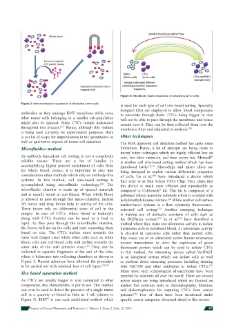
Figure 4: Microfl uidic-based separation of circulating tumor cells
            Figure 3: Immunomagnetic separation of circulating tumor cells
                                                              is used for such type of cell size based sorting. Specially
                                                              designed fi lter are employed to allow blood components
            antibodies as they undergo EMT transitions while some   to percolate through them. CTCs being bigger in size
            other tumor cells belonging to a smaller sub-population   will not be able to pass through the membrane and hence
            might also be ignored. Some CTCs remain undetected   remain over it. They can be then collected from over the
            throughout this process.  Hence, although this method   membrane fi lter and subjected to analysis. [32]
                                [27]
            is being used currently for experimental purposes, there
            is yet lot of scope for improvisation in the quantitative as   Other techniques
            well as qualitative aspects of tumor cell detection.  The FDA approved cell detection method has quite some
            Microfl uidics method                              limitations. Hence, a lot of attempts are being made to
                                                              invent better techniques which are highly effi cient low on
            As antibody-dependent cell sorting is not a completely   cost, less labor intensive, and time savers too. Metacell
                                                                                                            ®
            reliable source.  There are a lot of hurdles in   is another cell size-based sorting method which has been
            accomplishing higher percent enrichment of cells from   introduced lately. [32,33]  Microchips and micro slides are
            the whole blood. Hence, it is important to take into   being designed to exploit various differential properties
            consideration other methods which rely on antibody-free   of cells. Lu  et  al.  have introduced a device which
                                                                              [34]
            systems. In this method, cell size-based sorting is   they refer to as Nan Velcro CTCs Chip. They claim that
            accomplished using microfl uidic  technology. [28]  The   this device is much more effi cient and reproducible as
            microfl uidic chamber is made up of special materials   compared to CellSearch  kit.  This kit is composed of a
                                                                                  ®
            and is usually spiral or curvilinear.  When whole blood   patterned silicon nanowire substrate which is overlaid with
            is allowed to pass through this micro-chamber, inertial   polydimethylsiloxane mixture.  While another cell surface
                                                                                      [34]
            lift forces and drag forces help in sorting of the cells.   marker-based systems is a  fl ow  cytometry  fl uorescence-
            These forces rely on differential sizes of cell in the   activated cell sorting.  Another  emerging  technique
                                                                                 [35]
            sample. In case of CTCs, whole blood or leukocyte   is making use of dielectric constants of cells such as
            along with CTCs fraction can be used as a feed in   the DEPArray system.  Ju  et  al.  have described a
                                                                                            [36]
                                                                                 [26]
            input.  As they pass through the microfl uidic  chamber,   method where they make use telomerase activity to isolate
            the forces will act on the cells and start separating them   melanoma cells in peripheral blood. As telomerase activity
            based on size.  The CTCs incline more towards the   is elevated in cancerous cells rather than normal cells,
            inner wall (larger size) while other cells such as white   they made use of an adenoviral vector human telomerase
            blood cells and red blood cells will incline towards the   reverse transcriptase to drive the expression of green
            outer side of the wall (smaller size). [29]   They can be   fl uorescent protein which can be used to isolate CTCs
            collected in separate fragments at the end of the tube,   in this method.  An interesting device called  VeriFAST
            where it bifurcates into collecting chambers as shown in   is an integrated system which can isolate cells as well
            Figure 4. Recent advances have allowed the procedure   as perform down streaming processes including staining
            to be carried out with minimal loss of cell types. [30,31]  with EpCAM and other antibodies to isolate CTCs.
                                                                                                           [37]
            Size based separation method                      Many more such technological advancements have been
                                                              reported by scientists all over the world. There are several
            As CTCs are usually bigger in size compared to other   newer assays are being introduced which are focused on
            components, this characteristic is put to use. This method   marker free isolation such as chromatography,  fi ltration,
            can even be used to detect the presence of a single tumor   and dielectrophoresis for capturing CTCs from cancer
            cell in a quantity of blood as little as 1 mL (shown in   patients.  Few of them have been mentioned under
                                                                     [38]
                         ®
            Figure 5). ISET  is one such established method which   specifi c cancer categories discussed ahead in this review.
                Journal of Cancer Metastasis and Treatment  ¦  Volume 1 ¦ Issue 2 ¦ July 15, 2015 ¦        47