
Page 84 - Read Online
P. 84
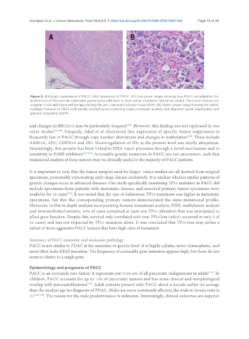
Skorupan et al. J Cancer Metastasis Treat 2023;9:5  https://dx.doi.org/10.20517/2394-4722.2022.106  Page 13 of 26


                Figure 3. Histologic appearance of PACC. H&E specimens of PACC. (A) Low power image showing how PACC recapitulates the
                architecture of the exocrine pancreatic parenchyma with back-to-back acinar structures containing lumina. The tumor clusters are
                irregular in size and shape and are approaching the peri- pancreatic adipose tissue (40×). (B) Higher power image showing the classic
                cytologic features of PACC with basally located nuclei containing single prominent nucleoli, and abundant apical amphophilic and
                granular cytoplasm (400×).


               and changes in BRCA1/2 may be particularly frequent . However, this finding was not replicated in two
                                                             [134]
               other studies [125,127] . Uniquely, Jakel et al. discovered that expression of specific tumor suppressors is
               frequently lost in PACC through copy number aberrations and changes in methylation . These include
                                                                                          [127]
               ARID1A, APC, CDKN2A and ID3. Downregulation of ID3 at the protein level was nearly ubiquitous.
               Interestingly, this protein has been linked to DNA repair processes through a novel mechanism and to
               sensitivity to PARP inhibitors [135-137] . Actionable genetic mutations in PACC are not uncommon, such that
               mutational analysis of these tumors may be clinically useful in the majority of PACC patients.

               It is important to note that the tumor samples used for larger -omics studies are all derived from surgical
               specimens, presumably representing early-stage disease exclusively. It is unclear whether similar patterns of
               genetic changes occur in advanced diseases. One study specifically examining TP53 mutation in PACC did
               include specimens from patients with metastatic disease, and matched primary tumor specimens were
               available for 10 cases . It was noted that the rate of deleterious TP53 mutations was higher in metastatic
                                 [138]
               specimens, but that the corresponding primary tumors demonstrated the same mutational profile.
               Moreover, in this in-depth analysis incorporating focused mutational analysis, FISH, methylation analysis
               and immunohistochemistry, 44% of cases contained at least one TP53 alteration that was anticipated to
               affect gene function. Despite this, survival only correlated with true TP53 loss (which occurred in only 5 of
               43 cases) and was not impacted by TP53 mutation alone. It was concluded that TP53 loss may define a
               subset of more aggressive PACC tumors that have high rates of metastasis.


               Summary of PACC anatomic and molecular pathology
               PACC is not similar to PDAC at the anatomic or genetic level. It is highly cellular, never desmoplastic, and
               most often lacks KRAS mutation. The frequency of actionable gene mutation appears high, but these do not
               seem to cluster to a single gene.

               Epidemiology and prognosis of PACC
                                                                                                     [139]
               PACC is an extremely rare tumor; it represents just 0.2%-2% of all pancreatic malignancies in adults . In
               children, PACC accounts for up to 15% of pancreatic tumors and has some clinical and morphological
                                           [119]
               overlap with pancreatoblastoma . Adult patients present with PACC about a decade earlier on average
               than the median age for diagnosis of PDAC. Males are more commonly affected; the male to female ratio is
               2:1 [140-143] . The reason for the male predominance is unknown. Interestingly, clinical outcomes are superior