
Page 21 - Read Online
P. 21
Dankbaar et al. J Cancer Metastasis Treat 2021;7:56 https://dx.doi.org/10.20517/2394-4722.2021.112 Page 5 of 8
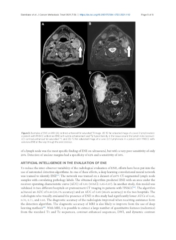
Figure 3. Examples of ENE on MR: (A) contrast-enhanced fat saturated T1 image; (B) T2 fat saturated image of a Level 2 lymph node in
a patient with HNSCC with minor ENE with subtle enhancement and T2 hyperintensity in the tissue around the lymph node (arrows);
(C) contrast-enhanced fat saturated T1; and (D) T2 fat saturated image of a Level 2/3 lymph node in a patient with HNSCC with
extensive ENE all the way through the skin (circles).
of a lymph node was the most specific finding of ENE on ultrasound, but with a very poor sensitivity of only
28%. Detection of unclear margins had a specificity of 62% and a sensitivity of 36%.
ARTIFICIAL INTELLIGENCE IN THE EVALUATION OF ENE
To reduce the inter-observer variability of the radiological evaluation of ENE, efforts have been put into the
use of automated detection algorithms. In one of these efforts, a deep learning convolutional neural network
[24]
was trained to identify ENE . The network was trained on a dataset of 2875 CT-segmented lymph node
samples with correlating pathology labels. The obtained algorithm predicted ENE with an area under the
receiver operating characteristic curve (AUC) of 0.91 (95%CI: 0.85-0.97). In another study, this model was
validated in two different hospitals on pretreatment CT imaging in patients with HNSCC . The algorithm
[40]
achieved an AUC of 0.84 (83.1% accuracy) and an AUC of 0.90 (88.6% accuracy) in the two hospitals. The
radiologists who visually evaluated the presence of ENE in this study had significantly lower AUCs of 0.60,
0.70, 0.71, and 0.82. The diagnostic accuracy of the radiologists improved when receiving assistance form
the detection algorithm. The diagnostic accuracy of MRI is also likely to improve from the use of deep
learning methods . With MRI, it is possible to extract a large number of quantitative features (radiomics)
[41]
from the standard T1 and T2 sequences, contrast enhanced sequences, DWI, and dynamic contrast