
Page 26 - Read Online
P. 26
Page 2 of 14 Woods et al. J Cancer Metastasis Treat 2022;8:22 https://dx.doi.org/10.20517/2394-4722.2022.28
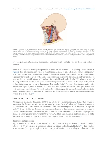
Figure 1. Assigned lymph node levels of the lateral neck. Level IA: Submental nodes. Level IB: Submandibular nodes. Level IIA: Upper
jugular nodes anterior to the spinal accessory nerve. Level IIB: Upper jugular nodes posterior to the spinal accessory nerve. Level III: Mid
jugular nodes. Level IV: Lower jugular nodes. Level VA: Posterior triangle nodes superior to the level of the inferior border of cricoid
cartilage. Level VB: Posterior triangle nodes inferior to the level of the inferior border of cricoid cartilage. Level VI: Central compartment
nodes.
pre- and post-auricular, parotid, suboccipital, and superficial lymphatic systems, depending on tumor
location.
Patterns of lymphatic drainage are predictable based on the location of the primary tumor, shown in
Figure 2. This information can be used to guide the management of regional diseases from specific primary
[1]
sites . As a general rule, a line joining the helix of one ear to the helix of the opposite ear in a coronal plane
separates the watershed areas of the scalp. Tumors located anterior to this line generally metastasize to
preauricular, periparotid, intraparotid, and anterior cervical lymph nodes (levels I-IV), whereas tumors of
the scalp posterior to this line usually metastasize to the postauricular and suboccipital lymph nodes, as well
as those in the posterior triangle of the neck and deep jugular chain . For example, cutaneous malignancies
[1]
on the cheek, eyelids, pinna, forehead, and temple have been reported as draining initially to preauricular,
[2]
periparotid, and parotid nodes . Most lymph nodes within the parotid are found superficial to the facial
nerve, and these are typically involved in cutaneous malignancy; however, a small number of nodes can be
present deep in the nerve .
[3,4]
RISK OF REGIONAL METASTASIS
Although non-melanoma skin cancer (NMSC) has a lower propensity for advanced disease than cutaneous
melanoma, the absolute mortality burden has recently surpassed that of melanoma . Cutaneous squamous
[5]
cell carcinoma (SCC) and Merkel cell carcinoma (MCC) have the highest risk of metastasis to regional
[6,7]
nodes . Other NMSCs can also present with regional diseases in the parotid and neck, such as cutaneous
adnexal tumors, cutaneous sarcomas and, rarely, basal cell carcinoma (BCC). The presence of lymph node
disease is a poor prognostic factor in NMSC. For SCC, it has been suggested that the presence of nodal
metastasis is a stronger predictor of prognosis than features present in the primary tumor .
[8]
Squamous cell carcinoma
Approximately 3.7%-5.8% of cases of cutaneous SCC present with regional disease [9-12] . However, higher
rates of 33%-47% are reported in association with several clinical and histological features [13,14] . These include
tumor location (ear, lip, or temple), size > 2 cm, depth of invasion > 6 mm or beyond subcutaneous fat,