
Page 23 - Read Online
P. 23
Page 4 of 12 Mansinho et al. J Cancer Metastasis Treat 2021;7:44 https://dx.doi.org/10.20517/2394-4722.2021.88
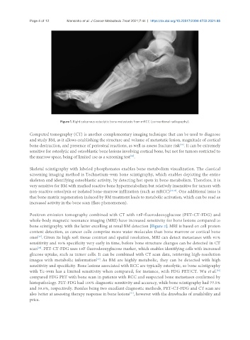
Figure 1. Right calcaneus osteolytic bone metastasis from mRCC (conventional radiography).
Computed tomography (CT) is another complementary imaging technique that can be used to diagnose
and study BM, as it allows establishing the structure and volume of metastatic lesion, magnitude of cortical
bone destruction, and presence of periosteal reactions, as well as assess fracture risk . It can be extremely
[33]
sensitive for osteolytic and osteoblastic bone lesions involving cortical bone, but not for tumors restricted to
the marrow space, being of limited use as a screening test .
[34]
Skeletal scintigraphy with labeled phosphonates enables bone metabolism visualization. The classical
screening imaging method is Technetium-99m bone scintigraphy, which enables depicting the entire
skeleton and identifying osteoblastic activity, by detecting hot spots in bone metabolism. Therefore, it is
very sensitive for BM with marked reactive bone hypermetabolism but relatively insensitive for tumors with
non-reactive osteolysis or isolated bone-marrow infiltration (such as mRCC) [35,36] . One additional issue is
that bone matrix regeneration induced by BM treatment leads to metabolic activation, which can be read as
increased activity in the bone scan (flare phenomenon).
Positron emission tomography combined with CT with 18F-fluorodeoxyglucose (PET-CT-FDG) and
whole-body magnetic resonance imaging (MRI) have increased sensitivity for bone lesions compared to
bone scintigraphy, with the latter excelling at renal BM detection [Figure 2]. MRI is based on cell proton
content detection, as cancer cells comprise more water molecules than bone marrow or cortical bone
[37]
ones . Given its high soft tissue contrast and spatial resolution, MRI can detect metastases with 91%
sensitivity and 95% specificity very early in time, before bone structure changes can be detected in CT
scan . PET-CT-FDG uses 18F-fluorodeoxyglucose marker, which enables identifying cells with increased
[38]
glucose uptake, such as tumor cells. It can be combined with CT scan data, retrieving high-resolution
images with metabolic information . As BM are highly metabolic, they can be detected with high
[39]
sensitivity and specificity. Bone lesions associated with RCC are typically osteolytic, so bone scintigraphy
with Tc-99m has a limited sensitivity when compared, for instance, with FDG PET/CT. Wu et al.
[40]
compared FDG PET with bone scan in patients with RCC and suspected bone metastases confirmed by
histopathology. PET-FDG had 100% diagnostic sensitivity and accuracy, while bone scintigraphy had 77.5%
and 59.6%, respectively. Besides being two excellent diagnostic methods, PET-CT-FDG and CT scan are
also better at assessing therapy response in bone lesions , however with the drawbacks of availability and
[41]
price.