
Page 22 - Read Online
P. 22
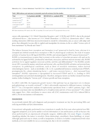
Mansinho et al. J Cancer Metastasis Treat 2021;7:44  https://dx.doi.org/10.20517/2394-4722.2021.88  Page 3 of 12

               Table 1. BM incidence and outcomes in metastatic renal cell carcinoma first-line studies
                Trial           % of patients with BM  HR (95%CI) vs. sunitinib (PFS)  HR (95%CI) vs. sunitinib (OS)
                Checkmate-214 [9]  20.0%            NR                          0.71 (0.47-1.08)
                          [10]
                Javelin Renal 101  NR               NR                          NR
                         [11]
                KEYNOTE-426     23.8%               NR                          NR
                           [12]
                CHECKMATE-9ER   24.1%               0.34 (0.22-0.55)            0.54 (0.32-0.92)
                    [13]
                CLEAR           23.9%               NR                          NR
               BM: Bone metastases; HR: hazard ratio; NR: not reported; OS: overall survival; PFS: progression-free survival.


               cancer cells expressing C-X-C Motif Chemokine Receptor 4 and 7 (CXCR4 and CXCR7), due to the stromal
                                                                                                   [23]
               cell-derived factor 1, also known as C-X-C Motif Chemokine 12 (CXCL12), chemotactic effect . After
               seeding, interaction with bone microenvironment is crucial for colonization, and, as cancer cells adapt and
               grow, they ultimately become able to exploit and manipulate the stroma, in the so-called “vicious cycle of
               bone metastases” by Mundy and Guise .
                                                [24]

               The balance between bone resorption and formation is well preserved in healthy bone, whereas it is
               disrupted and shifted towards bone resorption in BM. In physiological conditions, the triad of receptor
                                                                                                       [25]
               activator of nuclear factor kB ligand (RANKL), RANK, and osteoprotegerin (OPG) assures homeostasis .
               RANK is a transmembrane receptor that is present on the surface of osteoclast precursor cells and can be
               activated by its ligand RANKL, produced by osteoblasts, osteocytes, and bone marrow stromal cells. RANK
               activation by its ligand regulates osteoclast activity, survival, and differentiation . The RANKL soluble
                                                                                     [26]
               decoy OPG, secreted by mature osteoblasts and stromal cells, can block RANK activation and prevent bone
               resorption.  In  pathological  conditions,  such  as  those  of  osteolytic  metastases,  tumor-derived
               osteoclastogenic factors - such as interleukin 1 (IL-1), IL-6, parathyroid hormone-related protein (PTHrP),
               prostaglandin E2, colony-stimulating factor 1, and tumor necrosis factor-α - elicit increased bone
               resorption . RANKL  expression  is  upregulated  by  increased  PTHrP  and  IL-11,  leading  to  OPG
                        [27]
               downregulation and activated osteclastogenesis. Therefore, mitogenic factors are further released from bone
               matrix, further feeding the “vicious cycle” of tumor growth and bone resorption.


               In mRCC with BM, the hepatocyte growth factor/c-Met pathway has an important role in disease
               pathophysiology, as recently reviewed . c-Met is abnormally expressed in different tumors, including
                                                 [28]
               RCC [29-31] . In a retrospective analysis of nephrectomy specimens from 17 mRCC patients, high c-Met
                                                                                             [32]
               expression at primary sites was identified in 47% of nephrectomy and 86% of bone specimens . Pre-clinical
               data suggest that c-Met contributes to cancer stem cell (CSC) maintenance and CSCs-induced BM in
               mRCC, but its mechanism is still unknown.


               DIAGNOSIS
               As previously stated, BM early diagnosis and preemptive treatment are key for preventing SREs and
               reducing morbidity and QoL deterioration.


               Conventional radiography performed in two projections is usually the first exam when patients have BM-
               suggestive symptoms, as it can be useful to retrieve information on size and location, as well as fracture risk,
               when performed by an experienced orthopedic surgeon. Additionally, this exam [Figure 1] enables
               classifying whether the tumor is osteolytic (bone-destroying), osteoblastic (bone-building), or mixed.
               Although quick and easy to obtain, as well as inexpensive, it needs to be further complemented with other
               methods, as it is not suitable for use as a screening test, due to its low sensitivity.