
Page 7 - Read Online
P. 7
Nagayama. J Cancer Metastasis Treat 2021;7:6 I http://dx.doi.org/10.20517/2394-4722.2020.114 Page 3 of 16
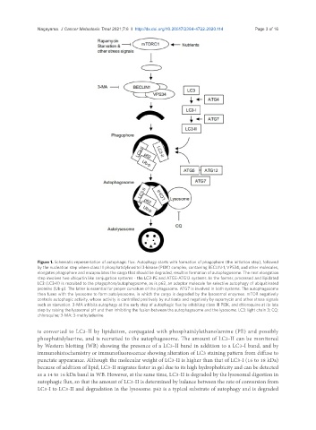
Figure 1. Schematic representation of autophagic flux. Autophagy starts with formation of phagophore (the initiation step), followed
by the nucleation step where class III phosphatidylinositol 3-kinase (PI3K) complex, containing BECLIN-1, VPS34, and other molecules,
elongates phagophore and encapsulates the cargo that should be degraded, result in formation of autophagosome. The next elongation
step involves two ubiquitin like conjugation systems - the LC3-PE and ATG5-ATG12 systems. In the former, processed and lipidated
LC3 (LC3-II) is recruited to the phagophore/autophagosome, as is p62, an adaptor molecule for selective autophagy of ubiquitinated
proteins (Ub-p). The latter is essential for proper curvature of the phagosome. ATG7 is involved in both systems. The autophagosome
then fuses with the lysosome to form autolysosome, in which the cargo is degraded by the lysosomal enzymes. mTOR negatively
controls autophagic activity, whose activity is controlled positively by nutrients and negatively by rapamycin and other stress signals
such as starvation. 3-MA inhibits autophagy at the early step of autophagic flux by inhibiting class III PI3K, and chloroquine at its late
step by raising the lysosomal pH and then inhibiting the fusion between the autophagosome and the lysosome. LC3: light chain 3; CQ:
chloroquine; 3-MA: 3-methyladenine
is converted to LC3-II by lipidation, conjugated with phosphatidylethanolamine (PE) and possibly
phosphatidylserine, and is recruited to the autophagosome. The amount of LC3-II can be monitored
by Western blotting (WB) showing the presence of a LC3-II band in addition to a LC3-I band, and by
immunohistochemistry or immunofluorescence showing alteration of LC3 staining pattern from diffuse to
punctate appearance. Although the molecular weight of LC3-II is higher than that of LC3-I (16 to 18 kDa)
because of addition of lipid, LC3-II migrates faster in gel due to its high hydrophobicity and can be detected
as a 14 to 16 kDa band in WB. However, at the same time, LC3-II is degraded by the lysosomal digestion in
autophagic flux, so that the amount of LC3-II is determined by balance between the rate of conversion from
LC3-I to LC3-II and degradation in the lysosome. p62 is a typical substrate of autophagy and is degraded