
Page 6 - Read Online
P. 6
Page 2 of 16 Nagayama. J Cancer Metastasis Treat 2021;7:6 I http://dx.doi.org/10.20517/2394-4722.2020.114
mutations in BRAF or RAS, or chromosomal rearrangements like RET/PTC as the causative role in
[2]
thyroid carcinogenesis . Thyroid cancer can be divided into two subgroups - differentiated [papillary
(PTC) and follicular (FTC)] and de-differentiated [poorly differentiated and anaplastic (ATC)] types.
PTC is the most common type, in which the mutant BRAF (BRAF V600E ) has been shown to be the most
[2]
frequent driver mutation, followed by RET/PTC and the mutant RAS . Surgery, radioactive iodine (RAI)
131
treatment with I and thyrotropin (TSH) suppression therapy have long been choices of treatment
modalities for thyroid cancer. In most patients with thyroid cancers, disease remission is achieved by these
modalities, but recurrence does occur in a subgroup of patients who acquired RAI resistance as a result
of dedifferentiation. Tyrosine kinase inhibitors have recently been approved as an additional therapeutic
choice for thyroid cancer treatment, especially for RAI refractory thyroid cancer, which clearly exhibit
its therapeutic benefits, but at the same time show some problems such as severe adverse effects and/or
intrinsic/acquired resistance. Thus, more novel therapeutic approach is urgently needed to be introduced.
Targeting autophagy may have a potential for this purpose.
Autophagy is an essential pathway mediating the degradation of cellular components such as proteins and
organelles in the lysosomes. It not only occurs constitutively at basal rate under physiological conditions
to maintain homeostasis for intracellular recycling of the metabolites and metabolic regulation, but it is
also induced during various physiological and pathological conditions such as nutritional starvation .
[3,4]
Alterations in autophagic activity have been widely considered to play crucial roles in numerous diseases
such as degenerative disorders, metabolic diseases, aging, and cancer, among others. There are three
types of autophagy so far identified in mammalian cells: macroautophagy (hereafter referred to as
“autophagy”), microautophagy, and chaperone-mediated autophagy (CMA). This review mainly focuses on
macroautophagy.
AUTOPHAGY IN PHYSIOLOGY
Autophagic flux
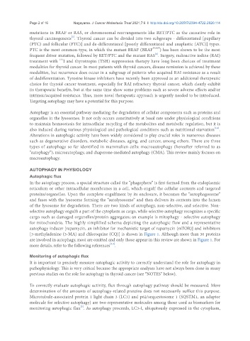
In the autophagy process, a special structure called the “phagophore” is first formed from the endoplasmic
reticulum or other intracellular membranes in a cell, which engulf the cellular contents and targeted
proteins/organelles. Upon the complete engulfment by its enclosure, it becomes the “autophagosome”
and fuses with the lysosome forming the “autolysosome” and then delivers its contents into the lumen
of the lysosome for degradation. There are two kinds of autophagy, non-selective, and selective. Non-
selective autophagy engulfs a part of the cytoplasm as cargo, while selective autophagy recognizes a specific
cargo such as damaged organelles/protein aggregates; an example is mitophagy - selective autophagy
for mitochondria. The highly simplified schema depicting the autophagic flow and a representative
autophagy inducer [rapamycin, an inhibitor for mechanistic target of rapamycin (mTOR)] and inhibitors
[3-methyladenine (3-MA) and chloroquine (CQ)] is shown in Figure 1. Although more than 30 proteins
are involved in autophagy, most are omitted and only those appear in this review are shown in Figure 1. For
[3,4]
more details, refer to the following references .
Monitoring of autophagic flux
It is important to precisely measure autophagic activity to correctly understand the role for autophagy in
pathophysiology. This is very critical because the appropriate analyses have not always been done in many
previous studies on the role for autophagy in thyroid cancer (see “NOTES” below).
To correctly evaluate autophagic activity, flux through autophagy pathway should be measured. Mere
determination of the amounts of autophagy-related proteins does not necessarily suffice this purpose.
Microtubule-associated protein 1 light chain 3 (LC3) and p62/sequestosome 1 (SQSTM1, an adaptor
molecule for selective autophagy) are two representative molecules among those used as biomarkers for
monitoring autophagic flux . As autophagy proceeds, LC3-I, ubiquitously expressed in the cytoplasm,
[5]