
Page 124 - Read Online
P. 124
Page 8 of 17 Priya et al. J Cancer Metastasis Treat 2021;7:70 https://dx.doi.org/10.20517/2394-4722.2021.122
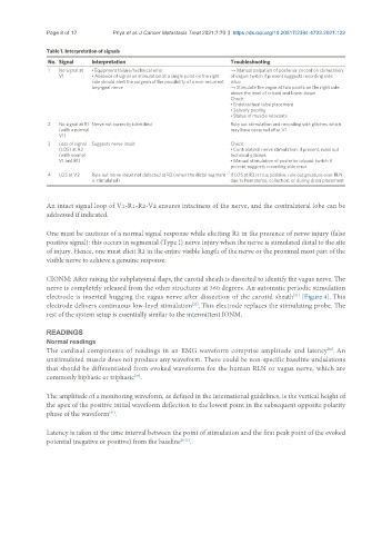
Table 1. Interpretation of signals
No. Signal Interpretation Troubleshooting
1 No signal at • Equipment failure/technical error → Manual palpation of posterior cricoid on stimulation
V1 • Absence of signal on stimulation at a single point on the right of vagus: twitch if present suggests recording side
side should alert the surgeon of the possibility of a non-recurrent issue
laryngeal nerve → Stimulate the vagus at two points on the right side:
above the level of cricoid and lower down
Check:
• Endotracheal tube placement
• Salivary pooling
• Status of muscle relaxants
2 No signal at R1 Nerve not correctly identified Rule out stimulation and recording side glitches which
(with a normal may have occurred after V1
V1)
3 Loss of signal Suggests nerve insult Check:
(LOS) at R2 • Contralateral nerve stimulation: if present, rules out
(with normal technical glitches
V1 and R1) • Manual stimulation of posterior cricoid: twitch if
present suggests recording side error
4 LOS at V2 Rule out nerve insult not detected at R2 (when the distal segment If LOS at R2 is true positive, rule out pressure over RLN
is stimulated) due to hematoma, collection, or during drain placement
An intact signal loop of V1-R1-R2-V2 ensures intactness of the nerve, and the contralateral lobe can be
addressed if indicated.
One must be cautious of a normal signal response while eliciting R2 in the presence of nerve injury (false
positive signal): this occurs in segmental (Type I) nerve injury when the nerve is stimulated distal to the site
of injury. Hence, one must elicit R2 in the entire visible length of the nerve or the proximal most part of the
visible nerve to achieve a genuine response.
CIONM: After raising the subplatysmal flaps, the carotid sheath is dissected to identify the vagus nerve. The
nerve is completely released from the other structures at 360 degrees. An automatic periodic stimulation
electrode is inserted hugging the vagus nerve after dissection of the carotid sheath [Figure 4]. This
[31]
[32]
electrode delivers continuous low-level stimulation . This electrode replaces the stimulating probe. The
rest of the system setup is essentially similar to the intermittent IONM.
READINGS
Normal readings
The cardinal components of readings in an EMG waveform comprise amplitude and latency . An
[36]
unstimulated muscle does not produce any waveform. There could be non-specific baseline undulations
that should be differentiated from evoked waveforms for the human RLN or vagus nerve, which are
commonly biphasic or triphasic .
[37]
The amplitude of a monitoring waveform, as defined in the international guidelines, is the vertical height of
the apex of the positive initial waveform deflection to the lowest point in the subsequent opposite polarity
[31]
phase of the waveform .
Latency is taken at the time interval between the point of stimulation and the first peak point of the evoked
potential (negative or positive) from the baseline [9,31] .