
Page 123 - Read Online
P. 123
Priya et al. J Cancer Metastasis Treat 2021;7:70 https://dx.doi.org/10.20517/2394-4722.2021.122 Page 7 of 17
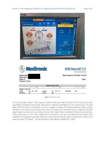
Figure 3. Electrode check.
recurrent laryngeal nerve . The surgery is further continued with retraction of the strap muscles and
[35]
exposing the ipsilateral thyroid lobe. Dissection is continued with ligation of the superior pole. The first
sight of the RLN either at the Beahr’s or Lorre’s triangle is confirmed with the monopolar probe, and the
waveform is recorded as the pre-dissection nerve (R1) signal. Once thyroidectomy is completed, the RLN
signal is again recorded as the post-dissection nerve (R2) signal. Here, stimulation of the most proximal part
of the exposed nerve or the entire visible nerve is necessary to rule out any segmental nerve injury. After
completion of hemostasis and approximation of the strap muscles, the vagus is stimulated to achieve Post
dissection vagal (V2) signal . The interpretation of the signals is tabulated in Table 1.
[35]