
Page 122 - Read Online
P. 122
Page 6 of 17 Priya et al. J Cancer Metastasis Treat 2021;7:70 https://dx.doi.org/10.20517/2394-4722.2021.122
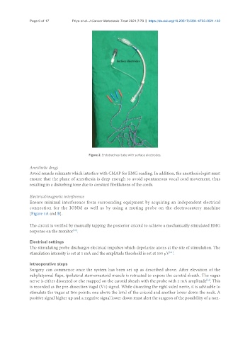
Figure 2. Endotracheal tube with surface electrodes.
Anesthetic drugs
Avoid muscle relaxants which interfere with CMAP for EMG reading. In addition, the anesthesiologist must
ensure that the plane of anesthesia is deep enough to avoid spontaneous vocal cord movement, thus
resulting in a disturbing tone due to constant fibrillations of the cords.
Electrical/magnetic interference
Ensure minimal interference from surrounding equipment by acquiring an independent electrical
connection for the IONM as well as by using a muting probe on the electrocautery machine
[Figure 1A and B].
The circuit is verified by manually tapping the posterior cricoid to achieve a mechanically stimulated EMG
[30]
response on the monitor .
Electrical settings
The stimulating probe discharges electrical impulses which depolarize axons at the site of stimulation. The
stimulation intensity is set at 1 mA and the amplitude threshold is set at 100 µV .
[31]
Intraoperative steps
Surgery can commence once the system has been set up as described above. After elevation of the
subplatysmal flaps, ipsilateral sternomastoid muscle is retracted to expose the carotid sheath. The vagus
nerve is either dissected or else mapped on the carotid sheath with the probe with 2 mA amplitude . This
[34]
is recorded as the pre-dissection vagal (V1) signal. While dissecting the right sided nerve, it is advisable to
stimulate the vagus at two points: one above the level of the cricoid and another lower down the neck. A
positive signal higher up and a negative signal lower down must alert the surgeon of the possibility of a non-