
Page 61 - Read Online
P. 61
Page 8 of 18 Malone et al. J Cancer Metastasis Treat 2021;7:40 https://dx.doi.org/10.20517/2394-4722.2021.37
Figure 2. Changes in gene expression and secretome in the brain parenchyma.
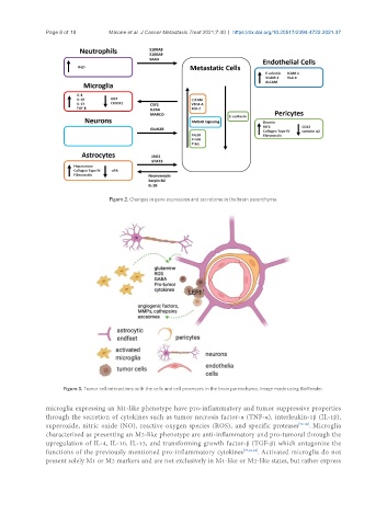
Figure 3. Tumor cell interactions with the cells and cell processes in the brain parenchyma. Image made using BioRender.
microglia expressing an M1-like phenotype have pro-inflammatory and tumor suppressive properties
through the secretion of cytokines such as tumor necrosis factor-α (TNF-α), interleukin-1β (IL-1β),
superoxide, nitric oxide (NO), reactive oxygen species (ROS), and specific proteases [78-80] . Microglia
characterized as presenting an M2-like phenotype are anti-inflammatory and pro-tumoral through the
upregulation of IL-4, IL-10, IL-13, and transforming growth factor-β (TGF-β) which antagonize the
functions of the previously mentioned pro-inflammatory cytokines [77,81,82] . Activated microglia do not
present solely M1 or M2 markers and are not exclusively in M1-like or M2-like states, but rather express