
Page 58 - Read Online
P. 58
Malone et al. J Cancer Metastasis Treat 2021;7:40 https://dx.doi.org/10.20517/2394-4722.2021.37 Page 5 of 18
[44]
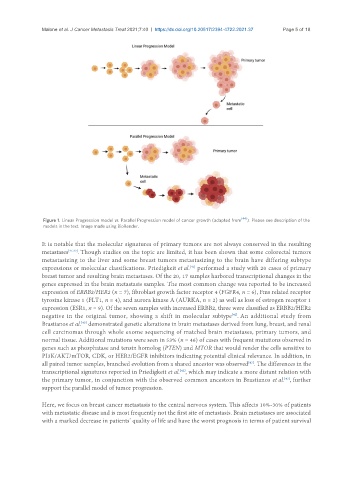
Figure 1. Linear Progression model vs. Parallel Progression model of cancer growth (adapted from ). Please see description of the
models in the text. Image made using BioRender.
It is notable that the molecular signatures of primary tumors are not always conserved in the resulting
metastases [41,42] . Though studies on the topic are limited, it has been shown that some colorectal tumors
metastasizing to the liver and some breast tumors metastasizing to the brain have differing subtype
[42]
expressions or molecular classifications. Priedigkeit et al. performed a study with 20 cases of primary
breast tumor and resulting brain metastases. Of the 20, 17 samples harbored transcriptional changes in the
genes expressed in the brain metastasis samples. The most common change was reported to be increased
expression of ERBB2/HER2 (n = 7), fibroblast growth factor receptor 4 (FGFR4, n = 6), Fms related receptor
tyrosine kinase 1 (FLT1, n = 4), and aurora kinase A (AURKA, n = 2) as well as loss of estrogen receptor 1
expression (ESR1, n = 9). Of the seven samples with increased ERBB2, three were classified as ERBB2/HER2
negative in the original tumor, showing a shift in molecular subtype . An additional study from
[42]
[43]
Brastianos et al. demonstrated genetic alterations in brain metastases derived from lung, breast, and renal
cell carcinomas through whole exome sequencing of matched brain metastases, primary tumors, and
normal tissue. Additional mutations were seen in 53% (n = 46) of cases with frequent mutations observed in
genes such as phosphatase and tensin homolog (PTEN) and MTOR that would render the cells sensitive to
PI3K/AKT/mTOR, CDK, or HER2/EGFR inhibitors indicating potential clinical relevance. In addition, in
all paired tumor samples, branched evolution from a shared ancestor was observed . The differences in the
[43]
transcriptional signatures reported in Priedigkeit et al. , which may indicate a more distant relation with
[42]
[43]
the primary tumor, in conjunction with the observed common ancestors in Brastianos et al. , further
support the parallel model of tumor progression.
Here, we focus on breast cancer metastasis to the central nervous system. This affects 10%-30% of patients
with metastatic disease and is most frequently not the first site of metastasis. Brain metastases are associated
with a marked decrease in patients’ quality of life and have the worst prognosis in terms of patient survival