
Page 24 - Read Online
P. 24
Page 6 of 20 Pellerino et al. J Cancer Metastasis Treat 2020;6:41 I http://dx.doi.org/10.20517/2394-4722.2020.80
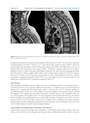
Figure 2. Diffuse linear spinal leptomeningeal enhancement from epidermal growth factor receptor and anaplastic lymphoma kinase wild-
type non-small-cell lung cancer
In general, the sensitivity of contrast-enhanced MRI in detecting LM is about 70%-85% with a specificity
[59]
[58]
of approximately 75%-90% . Freilich et al. have shown that contrast-enhanced MRI is altered in
approximately 90% of patients with LM from solid tumors and positive CSF, while about 20%-30% of
[4]
patients with LM may present a false-negative MRI . Therefore, a negative MRI does not exclude a
diagnosis of LM in a patient with typical neurological symptoms. No other alternative imaging techniques
have been used to validate negative MRI results in cases of high index of suspicion of LM. For instance,
there are no studies on the sensitivity and specificity of 18F-fluorodeoxyglucose positron emission
tomography–computed tomography (FDG-PET-CT) for LM diagnosis, due to resolution issues. In one
[60]
patient only a LM from NSCLC was detected using FDG-PET-CT .
CSF analysis
Some biochemical alterations may be found in the CSF of LM patients, such as an increased pressure (>
200 mm H O) in 21%-42% of patients, high level of proteins (> 50 mg/dL) in 56%-91%, decreased level
2
[61]
3
of glucose (<60 mg/dL), and elevated leucocyte count (> 4/mm ) in 48%-77.5% . All these findings are
not pathognomonic of LM, and a CSF cytology positive for neoplastic cells remains the gold standard
for diagnosis. CSF cytology does not allow a quantitative analysis and has a low sensitivity with 30%-
[57]
50% of LM patients with negative CSF . In case of negative results after the first CSF tap, the EANO/
ESMO Guidelines recommend performing a second lumbar puncture to improve the sensitivity up to 80%.
Moreover, there is no evidence that a conversion to a negative CSF is correlated with disease control in
leptomeninges and with a prolonged progression-free survival (PFS); thus, the cytological clearance of CSF
is not a reliable method to monitor leptomeningeal response.
Liquid biopsy in leptomeningeal metastases from NSCLC
Liquid biopsy consists of detecting tumor biomarkers in body fluids, such as blood, plasma, CSF, urine,
saliva, ascites, with the aim to diagnose and monitor disease. Different biomarkers may be detected,