
Page 12 - Read Online
P. 12
Su et al. J Cancer Metastasis Treat 2020;6:19 I http://dx.doi.org/10.20517/2394-4722.2020.48 Page 7 of 21
A B
C
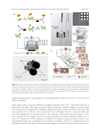
Figure 3. Schematic set-up for the microfluidic channel based immunomagnetic separation including enzyme catalysis and electrical
signal-off for detection of Salmonella (A) [43] (reprinted with permission from Elsevier); MACS tip for immunomagnetic separation
consisting of rectangular Ni meshes and permanent neodymium magnets on both sides of the Ni mesh (B) [46] (reprinted with
permission from Elsevier); schematic microfluidic channel design for the wash-free magnetic immunoassay for PSA using SERS droplet-
based sensor for readout (C) [47] (reprinted with permission from Royal Society of Chemistry). MACS: magnetic activated cell sorting;
PSA: prostate specific antigen; SERS: surface enhanced Raman scattering
significant improvement when compared to conventional devices where only 23.98% ± 6.59% of E. coli
could be separated.
[43]
Of the other works on using microfluidics for magnetic separation, Hou et al. reported the detection of
Salmonella from spiked milk using microfluidic signal off strategy to monitor changes in electrical signal.
This strategy helped in amplification of biological signals and the limit of detection (LOD) of the Salmonella
was reported to be 33 CFU/mL with as assay time of 2 h. Although it was not a rapid procedure, the
LOD seems promising. The schematic setup for their detection of Salmonella is shown in Figure 3A. The
mixture models for bio-magnetic separation using microfluidics were demonstrated by Khashan et al. .
[44]
[45]
Wu et al. reported size selective separation of magnetic microspheres using microfluidic channels. Their
[46]
experimental results were corroborated with numerical simulations. Oh et al. modified the tip of a
micropipette with Ni wire meshes and demonstrated magnetic activated cell sorting (MACS) [Figure 3B].