
Page 31 - Read Online
P. 31
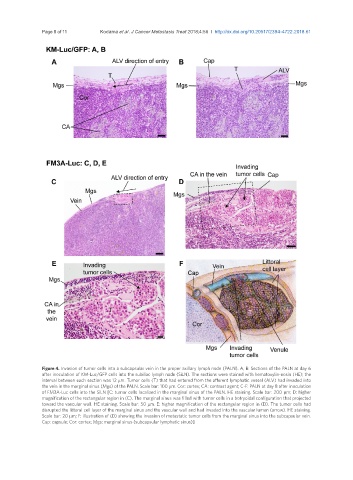
Page 8 of 11                          Kodama et al. J Cancer Metastasis Treat 2018;4:56  I  http://dx.doi.org/10.20517/2394-4722.2018.61


               Figure 4. Invasion of tumor cells into a subcapsular vein in the proper axillary lymph node (PALN). A, B: Sections of the PALN at day 6
               after inoculation of KM-Luc/GFP cells into the subiliac lymph node (SiLN). The sections were stained with hematoxylin-eosin (HE); the
               interval between each section was 12 μm. Tumor cells (T) that had entered from the afferent lymphatic vessel (ALV) had invaded into
               the vein in the marginal sinus (Mgs) of the PALN. Scale bar: 100 μm. Cor: cortex; CA: contrast agent; C-F: PALN at day 8 after inoculation
               of FM3A-Luc cells into the SiLN [C: tumor cells localized in the marginal sinus of the PALN. HE staining. Scale bar: 200 μm; D: higher
               magnification of the rectangular region in (C). The marginal sinus was filled with tumor cells in a botryoidal configuration that projected
               toward the vascular wall. HE staining. Scale bar: 50 μm. E: higher magnification of the rectangular region in (D). The tumor cells had
               disrupted the littoral cell layer of the marginal sinus and the vascular wall and had invaded into the vascular lumen (arrow). HE staining.
               Scale bar: 20 μm; F: illustration of (D) showing the invasion of metastatic tumor cells from the marginal sinus into the subcapsular vein.
               Cap: capsule; Cor: cortex; Mgs: marginal sinus (subcapsular lymphatic sinus)]