
Page 29 - Read Online
P. 29
Page 6 of 11 Kodama et al. J Cancer Metastasis Treat 2018;4:56 I http://dx.doi.org/10.20517/2394-4722.2018.61
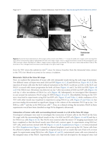
Figure 2. Flow in the blood vessel network of the proper axillary lymph node (PALN; n = 1 mouse, 12 weeks old). Images I and II are magnified
views of the corresponding regions highlighted in the left-most image. Image I shows a region that did not include the thoracoepigastric vein
(TEV) and was a frame obtained from Video 1. Image II shows a region that includes the TEV and was a frame obtained from Video 2. The
directions of flow in images I and II are shown schematically to the right of each image
[16]
from the TEV enters the subclavian vein ; since the venous branches from the intranodal veins connect
to the TEV, their blood is returned to the venous circulation.
Metastatic SiLN at the false N0 stage
Next, we explored the interaction of tumor cells with intranodal vessels during the early stage of metastasis.
Two different tumor cell types were used: KM-Luc/GFP [Figure 3A, C, E] and FM3A-Luc [Figure 3B, D, F]. The
injection of tumor cells into the SiLN induced metastasis in the PALN. Luciferase activity in the SiLN and
PALN increased with tumor progression for both cell lines (Figure 3A and C for KM-Luc/GFP, Figure 3B
and D for FM3A-Luc). Metastasis was detected on day 7 after inoculation of KM-Luc/GFP cells [Figure 3A]
and day 27 after inoculation of FM3A-Luc cells [Figure 3B]. Subsequently, we investigated flow dynamics
in and around the metastatic PALN using CE-HFUS [Figure 3E and F]. Microbubbles flowing in the TEV
were visualized in experiments using KM-Luc/GFP cells [Figure 3E and Video 3], while confluence of the
TEV with other vessels was visualized in experiments using FM3A-Luc cells [Figure 3F and Video 4]. Our
previous studies demonstrated no significant change in the volume of the metastatic PALN up to day 7 for
[20]
[21]
KM-Luc cells and day 22 for FM3A-Luc cells . Thus, in a clinical setting, the metastatic PALN in these
experiments would have been classified as stage N0 by diagnostic ultrasound.
Interaction of tumor cells with surrounding blood vessels in a LN at the false N0 stage
Histological techniques were used to investigate the interaction of tumor cells in the PALN (at the false
N0 stage) with the surrounding blood vessels at day 6 for KM-Luc/GFP cells [Figure 4A and B] and day 8
for FM3A-Luc cells [Figure 4C-E]. The PALNs were removed after the micro-CT imaging experiments had
been completed so that the blood vessels were filled with contrast agent. In experiments using KM-Luc/
GFP cells, serial sections stained with HE [Figure 4A and B, Supplementary Figure 1] or immunostained
for CD31 [Supplementary Figure 1] showed an afferent lymphatic vessel entering the LN. Tumor cells from
the afferent lymphatic vessel had invaded the marginal sinus as well as vessels that were filled with contrast
agent. In experiments using FM3A-Luc cells [Figure 4C and D], metastasized tumor cells were detected in
the marginal sinus of the PALN. Importantly, tumor cells that had developed in the marginal sinus had