
Page 213 - Read Online
P. 213
dos Santos et al. J Cancer Metastasis Treat 2019;5:25 I http://dx.doi.org/10.20517/2394-4722.2018.83 Page 5 of 20
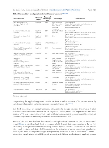
Table 1. Photosensitizers investigated in clinical trial for cancer treatment [15,21,23,30-35]
Treatment
Photosensitizer Chemical Wavelength Cancer type Characteristics
family
(nm)
Porfirmer sodium, HPD: Porphyrin 630 Lung, esophagus, 1st generation PS
hematoporphyrin derivative bile duct, bladder, Most probable intracellular localization: plasma
(Photofrin) brain, ovarian, membrane and mitochondria.
breast skin Intravenous administration
metastases
5-ALA: 5-aminolevulinic acid Porphyrin 630 Skin, bladder, brain, 2nd generation PS
(Levulan) precursor esophagus Most probable intracellular localization: mitochondria
Topical, oral or intravenous administration
MAL: methyl-aminolevulinate Porphyrin 630 Skin 2nd generation PS
(Metvix) precursor Most probable intracellular localization: mitochondria
and ER
Topical administration
h-ALA: hexylaminolevulinate Porphyrin White light Basal cell 2nd generation PS
(Hexvix) precursor Intracellular localization: TBD
Topical administration
Veteporfin, BDP: benzoporphyrin Porphyrin 690 Pancreas, breast 2nd generation PS
derivative (Visudyne) Most probable intracellular localization: mitochondria
Intravenous administration
Palladium bactereopheophorbide, Porphyrin 762 Esophagus, prostate 2nd generation PS
padeliporfin, WST-11 (Tookad) Intracellular localization: TBD. Intravenous
administration
Temoporfin, Chlorin 652 Head and neck, 2nd generation PS
mTHPC: meso- lung, brain, bile Most probable intracellular localization: mitochondria,
tetrahydroxyphenylchlorine duct, pancreas skin, golgi apparatus and ER
(Foscan) breast Intravenous administration
Talaporfin, mono-L-aspartyl chlorin Chlorin 660 Liver, colon, brain, 2nd generation PS
e6, NPe6, LS11 (Laserphyrin) lung, breast skin Most probable intracellular localization: lysosomes
metastases Intravenous administration
HPPH: Chlorin 665 Head and neck, 2nd generation PS
2-(1-hexyloxyethyl)-2-devinyl esophagus, lung Most probable intracellular localization: mitochondria
pyropheophorbide-a (Photochlor) and/or lysosomes
Intravenous administration
Rostaporfin, SnEt2: tin ethyl Chlorin 660 Skin, breast 2nd generation PS
etiopurpurin I, or (Purlytin) Most probable intracellular localization: lysosomes
Intravenous administration
Fimaporfin, disulfonated tetraphenyl Chlorin 633 Superficial cancers, 2nd generation PS
chlorin, TPCS2a (Amphinex) Cholon Most probable intracellular localization: endo-
lysosomal compartments
Intravenous administration
Motexafin lutetium (Lutex) Texaphyrin 732 Breast 2nd generation PS
Broad intracellular localization
Intravenous administration
TBD: to be determined
compromising the supply of oxygen and essential nutrients, as well as activation of the immune system, by
inducing an inflammatory and an immune response against tumor cells [23,35,40] .
Cell death subroutines are strongly connected with successful therapy outcome. Even when a detailed
explanation of cell death mechanisms is not the scope of this review (updated and deeper information can be
[39]
assessed in ), here we point some of their important features, since describing one or ones of them involved
in cell toxicity constitutes a very important topic of research in the field of PDT.
At the cellular level, PDT has been shown to induce multiple cell death subroutines, that can be accidental
or not [Figure 3]. Accidental cell death is an uncontrollable form of death corresponding to the physical
disassembly of the plasma membrane caused by extreme physical, chemical, or mechanical cues. On the
other hand, regulated cell death (RCD) results from the activation of one or more signal transduction
[39]
modules, and hence can be pharmacologically or genetically modulated, at least to some extent . The RCD
subroutines already related with PDT include apoptosis and different mechanisms of regulated necrosis