
Page 214 - Read Online
P. 214
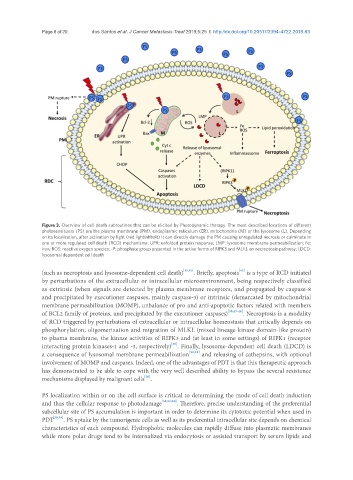
Page 6 of 20                   dos Santos et al. J Cancer Metastasis Treat 2019;5:25  I  http://dx.doi.org/10.20517/2394-4722.2018.83


               Figure 3. Overview of cell death subroutines that can be elicited by Photodynamic therapy. The most described locations of different
               photosensitizers (PS) are the plasma membrane (PM), endoplasmic reticulum (ER), mitochondria (M) or the lysosome (L). Depending
               on its localization, after activation by light (red lightinhbolt) it can directly damage the PM causing unregulated necrosis or culminate in
               one or more regulated cell death (RCD) mechanisms. UPR: unfolded protein response; LMP: lysosome membrane permeabilization; Fe:
               iron; ROS: reactive oxygen species; -P: phosphate group presented in the active forms of RIPK3 and MLKL on necroptosis pathway; LDCD:
               lysosomal dependent cell death

                                                                                  [42]
               (such as necroptosis and lysosome-dependent cell death) [15,41] . Briefly, apoptosis  is a type of RCD initiated
               by perturbations of the extracellular or intracellular microenvironment, being respectively classified
               as extrinsic (when signals are detected by plasma membrane receptors, and propagated by caspase-8
               and precipitated by executioner caspases, mainly caspase-3) or intrinsic (demarcated by mitochondrial
               membrane permeabilization (MOMP), unbalance of pro and anti-apoptotic factors related with members
               of BCL2 family of proteins, and precipitated by the executioner caspases) [39,43-48] . Necroptosis is a modality
               of RCD triggered by perturbations of extracellular or intracellular homeostasis that critically depends on
               phosphorylation, oligomerization and migration of MLKL (mixed lineage kinase domain-like protein)
               to plasma membrane, the kinase activities of RIPK3 and (at least in some settings) of RIPK1 (receptor
                                                           [49]
               interacting protein kinases-1 and -3, respectively) . Finally, lysosome-dependent cell death (LDCD) is
               a consequence of lysosomal membrane permeabilization [50,51]  and releasing of cathepsins, with optional
               involvement of MOMP and caspases. Indeed, one of the advantages of PDT is that this therapeutic approach
               has demonstrated to be able to cope with the very well described ability to bypass the several resistance
                                                  [26]
               mechanisms displayed by malignant cells .

               PS localization within or on the cell surface is critical to determining the mode of cell death induction
               and thus the cellular response to photodamage [38,52,53] . Therefore, precise understanding of the preferential
               subcellular site of PS accumulation is important in order to determine its cytotoxic potential when used in
               PDT [38,54] . PS uptake by the tumorigenic cells as well as its preferential intracellular site depends on chemical
               characteristics of each compound. Hydrophobic molecules can rapidly diffuse into plasmatic membranes
               while more polar drugs tend to be internalized via endocytosis or assisted transport by serum lipids and