
Page 58 - Read Online
P. 58
Enrique et al. J Cancer Metastasis Treat 2019;5:54 I http://dx.doi.org/10.20517/2394-4722.2019.20 Page 3 of 16
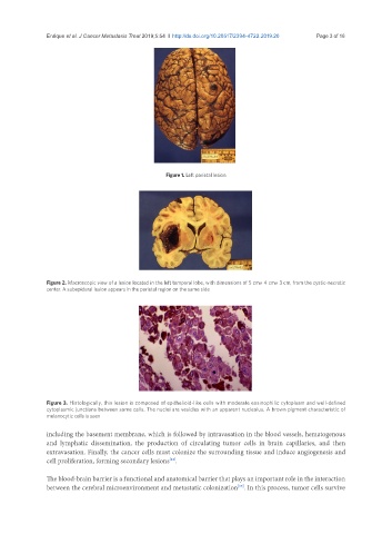
Figure 1. Left parietal lesion
Figure 2. Macroscopic view of a lesion located in the left temporal lobe, with dimensions of 5 cm× 4 cm× 3 cm, from the cystic-necrotic
center. A subepidural lesion appears in the parietal region on the same side
Figure 3. Histologically, this lesion is composed of epithelioid-like cells with moderate eosinophilic cytoplasm and well-defined
cytoplasmic junctions between some cells. The nuclei are vesicles with an apparent nucleolus. A brown pigment characteristic of
melanocytic cells is seen
including the basement membrane, which is followed by intravasation in the blood vessels, hematogenous
and lymphatic dissemination, the production of circulating tumor cells in brain capillaries, and then
extravasation. Finally, the cancer cells must colonize the surrounding tissue and induce angiogenesis and
cell proliferation, forming secondary lesions .
[14]
The blood-brain barrier is a functional and anatomical barrier that plays an important role in the interaction
between the cerebral microenvironment and metastatic colonization . In this process, tumor cells survive
[15]