
Page 60 - Read Online
P. 60
Enrique et al. J Cancer Metastasis Treat 2019;5:54 I http://dx.doi.org/10.20517/2394-4722.2019.20 Page 5 of 16
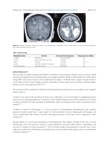
Figure 4. Magnetic resonance imaging of a patient with multiple brain metastases. Axial T2 slice sequence shows perilesional edema in
the junction of white and gray matter
Table 1. Spectroscopy
Metabolite/marker Function As found in brain metastases Range (parts per million)
Creatine Metabolism Internal standard 3.0
Choline Cellular membrane turnover Increased 3.2
Lipids Necrosis Increased 0.9-1.4
Lactate Anaerobic metabolism/necrosis Increased 1.3
N-acetylaspartate Neuronal viability Decreased 2.0
SPECTROSCOPY
Spectroscopy is an MRI technique that allows the metabolic characteristics of lesions to be evaluated, which
may help distinguish between benign lesions and malignant lesions which would be difficult to differentiate
using MRI alone. Spectroscopy can be performed for single or multiple tumor regions (unique voxel or
multivoxel) to detect certain ranges of specific metabolites in brain tissue, such as choline, creatinine, lipids,
lactate, and N-acetyl-aspartate (NAA) [22,23] .
The analysis of these metabolites is helpful for distinguishing metastasis from necrosis, gliosis, and vasogenic
edema [Table 1].
Creatine is the most stable metabolite in brain tissue, although it can be diminished in malignant primary
tumors such as high-grade gliomas. It is present in both white and gray matter, which enables it to be used as
an internal reference for the remainder of metabolites, which can change in the context of metastatic brain
disease [24,25] .
Choline is a marker of cell change: it is a structural part of cell membrane phospholipids, with a greater
presence in white matter than in gray matter. It is elevated where there are high-grade cell changes, and
it has a relationship with creatine, such that both appear elevated, which helps orient a diagnosis of brain
metastasis .
[26]
Because lipids are a structural component of cell membranes, they appear elevated in the case of severe
cell damage, even with necrosis. Lactate is a main metabolite associated with anaerobic metabolism, and
necrosis is common in brain metastases . NAA is found at high concentrations in normal brain tissue,
[26]
making it a marker for cell integrity and normal tissue structure; this marker appears at low concentrations
in brain metastases [25,26] .