
Page 38 - Read Online
P. 38
Page 4 of 13 Yagishita et al. J Cancer Metastasis Treat 2019;5:75 I http://dx.doi.org/10.20517/2394-4722.2019.026
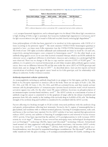
Table 2. Characteristics of IgG isotypes
Heavy chain subtype Isotype ADCC activity CDC activity Half-life (days)
g1 IgG1 ++ ++ 21
g2 IgG2 + + 20
g3 IgG3 +++ +++ 7
g4 IgG4 + - 21
ADCC: antibody-dependent cellular cytotoxicity; CDC: complement-derived cellular cytotoxicity
< 6.0), escapes lysosomal degradation, and is released again into the blood. If the blood IgG concentration
is high, binding of FcRn to IgG is saturated, then lysosome-mediated IgG degradation is enhanced, and if
[10]
the IgG concentration is low, IgG is bound to FcRn and recycled, thereby reducing IgG degradation .
Gene polymorphism of FcRn has been reported to be involved in FcRn expression, with VNTR of 37
[11]
bases occurring in the promoter region . The most common VNTR3/VNTR3 homozygous genotype is
[11]
reported to have 1.66 times more FcRn expression than the VNTR3/VNTR2 heterozygous genotype .
The levels of anti-TNF-alpha antibodies infliximab and adalimumab are lower in blood by 14% and 24%,
[12]
respectively, among heterozygous cases compared to homozygous cases . On the other hand, as an
antibody drug against cancer, in 94 cases that received cetuximab therapy, which is an EGFR antibody for
colorectal cancer, a significant difference in distribution clearance and a tendency to a prolonged half-life
[13]
were observed. There was no change in PK due to copy number variation (CNV) of FCGRT gene . In
addition, in 476 patients who received farletuzumab, an anti-folate receptor alpha antibody against ovarian
cancer, there was no difference between PK and the area under the curve (AUC) of VNTR genotype and
[14]
steady-state, and no change in PK due to CNV . As described above, there are few reports on blood
concentrations of antibody drugs against cancer based on the FcRn genotype, and the relationship with
efficacy is unknown. Further evidence is needed.
Antibody-dependent cellular cytotoxicity
An immunoglobulin including an antibody drug binds to an antigen at the Fab region, and the Fc region
binds to Fcg Receptor expressed on immune cells in the body. There are six types of FcgR: I, IIA, IIB,
IIC, IIIA, and IIIB. Of them, I, IIA, IIC, IIIA, and IIIB are activated forms, and they mainly activate
immune cells by phosphorylation of immunoreceptor tyrosine-based activation motif, which transmits
activation signals into cells. On the other hand, IIB causes inhibitory functions via phosphorylation of
[15]
an immunoreceptor tyrosine-based inhibitory motif . The ADCC activity as a mechanism of action of
antibody drugs for cancer is considered to be the apoptosis of target cells by the release of perforin and
granzyme from NK cells, mainly by the binding of the antibody drug to FcgRIIIA of NK cells. The binding
strength of the antibody drug and FcgRIIIA is considered to be correlated with the ADCC activity.
Factors affecting the binding strength to FCgR include structural problems with the antibody drug
and genetic polymorphisms affecting host immunity. Normally, the Fc region of immunoglobulins is
glycosylated, and variations with or without terminal galactose, bisecting N-acetylglucosamine, sialic acid,
and fucose at the root have been reported. For the ADCC activity, it has been reported that aglycosylation
reduces ADCC activity, and addition of bisecting N-acetylglucosamine and removal of fucose increase
ADCC activity. It has been reported that the addition of N-acetylglucosamine to mAb increases ADCC
[16]
activity by 10-20 times . Moreover, fucose removal has undergone many technological developments,
TM
TM
such as Potelligent technology (BioWa, Japan) and Glycomab technology (Glycart, Switzerland)
based around cell lines engineered with altered glycosylation machinery, that increase ADCC activity
TM
by more than 100-fold [17,18] . In the Potelligent cell line, both FUT8 gene alleles, which encode a1,6-
TM
fucosyltransferase, were disrupted by sequential homologous recombination. The Glycomab cell lines are
stably transfected with the gene encoding 1,4-N-acetylglucosaminyltransferse III (GnIII), resulting in the