
Page 856 - Read Online
P. 856
Silk et al. Hepatoma Res 2020;6:73 I http://dx.doi.org/10.20517/2394-5079.2020.61 Page 7 of 16
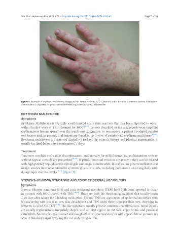
Figure 5. Example of erythema multiforme. Image author James Heilman, MD. Obtained under Creative Commons license Attribution-
Share Alike 3.0 Unported. https://creativecommons.org/licenses/by-sa/3.0/deed.en
ERYTHEMA MULTIFORME
Symptoms
Erythema Multiforme is typically a self-limited acute skin reaction that has been reported to occur
within the first week of TKI treatment for HCC [40-42] . Lesions described in the case reports were targetoid
erythematous lesions spread over the trunk and extremities. In one report, a patient developed painful
oral lesions and, in general, oral lesions are found in up to 60% of people with erythema multiforme [42,43] .
Erythema multiforme is diagnosed clinically based on the patient’s history and physical examination. It
usually has fixed lesions for a minimum of 7 days.
Treatment
Treatment involves medication discontinuation. Additionally for mild disease oral antihistamines with or
without topical steroids are prescribed [42,44] . If painful mucosal erosions are present, they can be treated
with high potency topical corticosteroid gels and magic mouthwashes. If oral lesions prevent sufficient oral
intake, studies have recommended systemic glucocorticoids, including prednisone 40-60 mg daily with
dosage taper over 2-4 weeks [42,44] [Figure 5].
STEVENS-JOHNSON SYNDROME AND TOXIC EPIDERMAL NECROLYSIS
Symptoms
Stevens-Johnson syndrome (SJS) and toxic epidermal necrolysis (TEN) have both been reported to occur
in patients with HCC treated with TKIs [40,45] . These are both life-threatening reactions that usually begin
4-28 days after taking the offending medication. SJS and TEN are a spectrum of epidermal necrolysis with
SJS occurring with less than 10% skin detachment and TEN when there is greater than 30%. Anything in
between is called SJS-TEN [46,47] . Flu-like symptoms usually precede cutaneous manifestations. Initial lesions
are usually erythematous, irregularly shaped, and can first appear on the face, upper trunk, and proximal
extremities. Necrotic lesions coalesce and slough off either spontaneously or with applied lateral pressure (as
seen in Nikolsky’s sign) revealing the red underlying dermis.