
Page 765 - Read Online
P. 765
Shimizuguchi et al. Hepatoma Res 2020;6:66 I http://dx.doi.org/10.20517/2394-5079.2020.51 Page 3 of 10
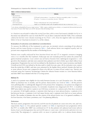
Table 1. Criteria of adverse factors
Factor Criteria
1. Physical factor
General condition ECOG performance status = 2 or above or Charlson comorbidity index = 5 or above
Liver function CP score = 7 or above or normal liver volume < 1000 mL*
2. Tumor factor
Tumor persistence Three or more previous liver tumor treatments**
Planning difficulty Target is in immediate contact with the gastrointestinal tract or tumor size > 5 cm
*Liver volume subtracted by the gross target volume; **either surgery, radiofrequency ablation, or transarterial chemoembolization for
liver tumor. CP score: Child-Pugh score; ECOG: European Clinical Oncology Group
in 5 fractions was selected to reduce the normal liver dose, while a more fractionated schedule (40 Gy in 10
fractions) was selected for cases in which the PTV was in direct contacted with the OARs. Dose constraint
criteria for the liver were volumes receiving 20 Gy (V20) < 20%. Dose for digestive tube was restricted
below 30 Gy in 5 fractions to at most 0.5 cc of the volume.
Evaluation of outcomes and statistical considerations
To measure the difficulty of the treatment in each case, we invented criteria consisting of two physical
factors and two tumor factors as shown in Table 1. Each adverse factor was weighted equally, and the
patients were categorized by the number of harboring factors.
Patients were usually evaluated by liver function blood test and CT or magnetic resonance imaging
every 3 to 6 months after the treatment. Endoscopy was not routinely performed unless the patient had
gastrointestinal symptoms. Local control was defined as freedom from radiological progression (> 20%
growth in the diameter), and data were censored when patients were lost to follow-up or died without local
progression. Progression-free survival was defined as the duration to any liver tumor recurrence and death.
Overall survival (OS) was defined as the time until to death from any cause. All the indicators were counted
from the initial day of SBRT, and rates were estimated by the Kaplan-Meier method. Patients who had two
or more adverse factors were compared to those who did not, in terms of local control rate. Toxicity was
evaluated using the Common Terminology Criteria for Adverse Events version 4.0. Liver function before
and after SBRT was evaluated with the CP scoring system.
RESULTS
A total of 24 patients were eligible for this study between January 2014 and December 2019. The median
follow-up duration was 18 months, and the patient characteristics are listed in Table 2. All the patients
were pathologically or radiologically diagnosed with primary liver malignancy; one patient was diagnosed
with intrahepatic cholangiocarcinoma, the remainder of patients were diagnosed with HCC. A modified
SBRT prescription was used in 9 patients (an OAR dose constraint in 3 patients and a liver dose constraint
in 6 patients). Of all eligible patients, 21 patients (88%) had one or more adverse factors, 16 patients (67%)
had physical factors, 13 patients (54%) had tumor factors, and 11 patients (46%) had two or more adverse
factors. The number of patients who met the criteria of the adverse factor was 10 for general condition, 10
for liver function, 9 for tumor persistence, and 7 for planning difficulty.
Outcomes
The local control, progression-free survival, and OS rates for all patients at 2 years were 89%, 42%, and 76%,
respectively [Figure 1]. One patient (patient number 8 in Table 2) experienced local progression within
the PTV 20 months after SBRT was confirmed on CT. In the patients with physical and tumor adverse
factors, local control/progression-free survival/OS rates at 2 years were 100%/42%/69% and 80%/23%/78%,
respectively. The subgroup of 11 patients with 2 or more (13 patients with 0 to 1) adverse factors showed