
Page 18 - Read Online
P. 18
Page 6 of 10 Umetsu et al. Hepatoma Res 2020;6:1 I http://dx.doi.org/10.20517/2394-5079.2019.030
A B
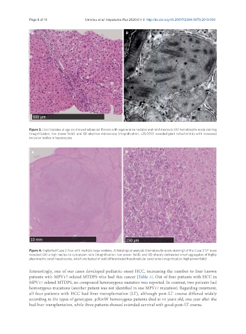
Figure 3. Liver biopsies at age six showed advanced fibrosis with regenerative nodules and mild steatosis: (A) hematoxylin-eosin staining
(magnification, low power field); and (B) electron microscopy (magnification, ×25,000) revealed giant mitochondria with increased
inclusion bodies in hepatocytes
A B
Figure 4. Explanted Case 2 liver with multiple large nodules. A histological analysis (hematoxylin-eosin staining) of the Case 2 S7 mass
revealed: (A) a high nucleo-to-cytoplasm ratio (magnification, low power field); and (B) sharply delineated small aggregates of highly
pleomorphic small hepatocytes, which are typical of well-differentiated hepatocellular carcinoma (magnification, high power field)
Interestingly, one of our cases developed pediatric-onset HCC, increasing the number to four known
patients with MPV17-related MTDPS who had this cancer [Table 3]. Out of four patients with HCC in
MPV17-related MTDPS, no compound heterozygous mutation was reported. In contrast, two patients had
homozygous mutations (another patient was not identified in one MPV17 mutation). Regarding treatment,
all four patients with HCC had liver transplantation (LT), although post-LT course differed widely
according to the types of genotypes. p.R50W homozygous patients died at 10 years old, one year after she
had liver transplantation, while three patients showed extended survival with good-post-LT course.