
Page 16 - Read Online
P. 16
Page 4 of 10 Umetsu et al. Hepatoma Res 2020;6:1 I http://dx.doi.org/10.20517/2394-5079.2019.030
A B
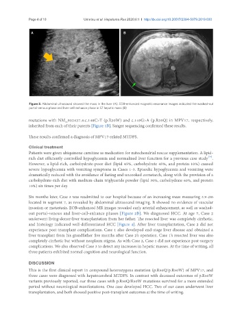
Figure 2. Abdominal ultrasound showed the mass in the liver (A). EOB-enhanced magnetic resonance images indicated the washed-out
portal-venous phase and liver-cell-enhance phase in S7 hepatic mass (B)
mutations with NM_002437.4:c.148C>T (p.R50W) and c.149G>A (p.R50Q) in MPV17, respectively,
inherited from each of their parents [Figure 1B]. Sanger sequencing confirmed these results.
These results confirmed a diagnosis of MPV17-related MTDPS.
Clinical treatment
Patients were given ubiquinone carnitine as medication for mitochondrial rescue supplementation. A lipid-
[14]
rich diet efficiently controlled hypoglycemia and normalized liver function for a previous case study .
However, a lipid-rich, carbohydrate-poor diet (lipid 45%, carbohydrate 45%, and protein 10%) caused
severe hypoglycemia with vomiting symptoms in Cases 1-3. Episodic hypoglycemia and vomiting were
dramatically reduced with the avoidance of fasting and uncooked cornstarch, along with the provision of a
carbohydrate-rich diet with medium-chain triglyceride powder (lipid 30%, carbohydrate 60%, and protein
10%) six times per day.
Six months later, Case 2 was readmitted to our hospital because of an increasing mass measuring 2.9 cm
located in segment 7, as revealed by abdominal ultrasound imaging. It showed no evidence of vascular
invasion or metastasis. EOB-enhanced MR images revealed early arterial enhancement, as well as washed-
out portal-venous and liver-cell-enhance phases [Figure 2B]. We diagnosed HCC. At age 7, Case 2
underwent living-donor-liver transplantation from her father. The resected liver was completely cirrhotic,
and histology indicated well-differentiated HCC [Figure 4]. After liver transplantation, Case 2 did not
experience post-transplant complications. Case 1 also developed end-stage liver disease and obtained a
liver transplant from his grandfather five months after Case 2’s operation. Case 1’s resected liver was also
completely cirrhotic but without neoplasm stigma. As with Case 2, Case 1 did not experience post-surgery
complications. We also observed Case 3 to detect any increases in hepatic masses. At the time of writing, all
three patients exhibited normal cognition and neurological function.
DISCUSSION
This is the first clinical report in compound heterozygous mutation (p.R50Q/p.R50W) of MPV17, and
three cases were diagnosed with hepatocerebral MTDPS. In contrast with deceased outcomes of p.R50W
variants previously reported, our three cases with p.R50Q/R50W mutations survived for a more extended
period without neurological manifestations. One case developed HCC. Two of our cases underwent liver
transplantation, and both showed positive post-transplant outcomes at the time of writing.