
Page 15 - Read Online
P. 15
Umetsu et al. Hepatoma Res 2020;6:1 I http://dx.doi.org/10.20517/2394-5079.2019.030 Page 3 of 10
A B
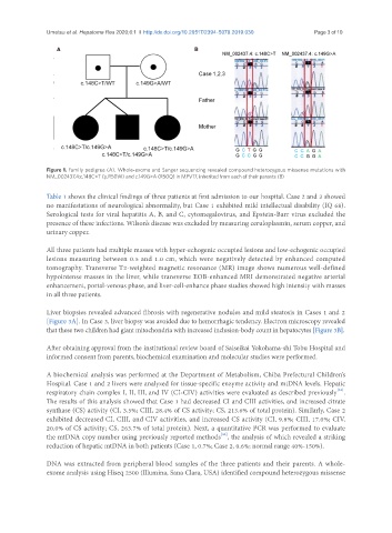
Figure 1. Family pedigree (A). Whole-exome and Sanger sequencing revealed compound heterozygous missense mutations with
NM_002437.4:c.148C>T (p.R50W) and c.149G>A (R50Q) in MPV17, inherited from each of their parents (B)
Table 1 shows the clinical findings of three patients at first admission to our hospital. Case 2 and 3 showed
no manifestations of neurological abnormality, but Case 1 exhibited mild intellectual disability (IQ 68).
Serological tests for viral hepatitis A, B, and C, cytomegalovirus, and Epstein-Barr virus excluded the
presence of these infections. Wilson’s disease was excluded by measuring ceruloplasmin, serum copper, and
urinary copper.
All three patients had multiple masses with hyper-echogenic occupied lesions and low-echogenic occupied
lesions measuring between 0.5 and 1.0 cm, which were negatively detected by enhanced computed
tomography. Transverse T2-weighted magnetic resonance (MR) image shows numerous well-defined
hypointense masses in the liver, while transverse EOB-enhanced MRI demonstrated negative arterial
enhancement, portal-venous phase, and liver-cell-enhance phase studies showed high intensity with masses
in all three patients.
Liver biopsies revealed advanced fibrosis with regenerative nodules and mild steatosis in Cases 1 and 2
[Figure 3A]. In Case 3, liver biopsy was avoided due to hemorrhagic tendency. Electron microscopy revealed
that these two children had giant mitochondria with increased inclusion-body count in hepatocytes [Figure 3B].
After obtaining approval from the institutional review board of Saiseikai Yokohama-shi Tobu Hospital and
informed consent from parents, biochemical examination and molecular studies were performed.
A biochemical analysis was performed at the Department of Metabolism, Chiba Prefectural Children’s
Hospital. Case 1 and 2 livers were analyzed for tissue-specific enzyme activity and mtDNA levels. Hepatic
[31]
respiratory chain complex I, II, III, and IV (CI-CIV) activities were evaluated as described previously .
The results of this analysis showed that Case 1 had decreased CI and CIII activities, and increased citrate
synthase (CS) activity (CI, 3.5%; CIII, 28.4% of CS activity; CS, 213.6% of total protein). Similarly, Case 2
exhibited decreased CI, CIII, and CIV activities, and increased CS activity (CI, 9.8%; CIII, 17.0%; CIV,
20.0% of CS activity; CS, 263.7% of total protein). Next, a quantitative PCR was performed to evaluate
[32]
the mtDNA copy number using previously reported methods , the analysis of which revealed a striking
reduction of hepatic mtDNA in both patients (Case 1, 0.7%; Case 2, 0.6%; normal range 40%-150%).
DNA was extracted from peripheral blood samples of the three patients and their parents. A whole-
exome analysis using Hiseq 2500 (Illumina, Sana Clara, USA) identified compound heterozygous missense