
Page 192 - Read Online
P. 192
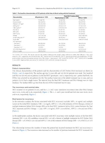
Page 4 of 11                                          Nakamura et al. Hepatoma Res 2019;5:16  I  http://dx.doi.org/10.20517/2394-5079.2019.06


               Table 1. The baseline characteristics of 312 patients at the time of direct-acting antiviral treatment
                                                                          Final HCC Tx before DAA
                Characteristics          All patients (n = 312)
                                                               Hepatectomy (n = 89)       RFA (n = 223)
                Age (years)                 74 (68-79)              74 (68-78)            74 (68-79)
                Gender (male)               182 (58.3%)             50 (56.2%)            132 (59.1%)
                        3
                Platelet (× 10 /μL)*        105 (76-140)            113 (98-137)          99 (73-142)
                ALB (g/dL)*                 3.8 (3.4-4.0)           3.9 (3.6-4.2)         3.7 (3.4-4.0)
                T.Bil (mg/dL)**             0.8 (0.6-1.1)           0.73 (0.6-0.9)        0.89 (0.6-1.2)
                AST (U/L)                   52 (37-67)              51 (39-70)            52 (36-66)
                ALT (U/L)                   41 (28-60)              42 (31-65)            39 (27-60)
                GGTP (U/L)                  37 (25-55)              40 (26-62)            36 (24-52)
                PT-INR                      1.07 (1.0-1.14)         1.07 (0.99-1.11)      1.06 (1.00-1.15)
                Child-Pugh grade A**        280 (89.7%)             83 (93.3%)            197 (88.3%)
                Fib-4 index                 5.64 (3.75-8.04)        5.06 (3.65-6.85)      5.88 (3.75-8.53)
                HCV genotype*
                   Genotype 1               251 (80.5%)             65 (73.0%)            186 (83.4%)
                   Genotype 2               60 (19.2%)              24 (27.0%)            36 (16.2%)
                   Genotype 1 + 2           1 (0.3%)                0 (0%)                1 (0.4%)
                HCV-RNA (log IU/mL)         6.0 (5.4-6.4)           6.0 (5.4-6.5)         6.0 (5.4-6.4)
                SVR at 12 weeks             288 (92.3%)             83 (93.3%)            205 (91.9%)
               *P < 0.001; **P < 0.05; The values indicate the median (interquartile range) unless otherwise noted. ALB: albumin; T.Bil: total
               bilirubin; AST: aspartate aminotransferase; ALT: alanine aminotransferase; GGTP: gamma-glutamyl transpeptidase; PT-INR:
                                                                                         1/2
                                                                                  9
               prothrombin time-international normalized ratio; FIB-4 index = Age (years) × AST (U/L)/[PLT(10 /L) × ALT  (U/L)]; HCV: hepatitis
               C virus; HCC: hepatocellular carcinoma; Tx: treatment; SVR: sustained virological response
               RESULTS
               Patient characteristics
               The clinical characteristics of the patients and the characteristics of HCC before DAA treatment are shown in
               [Tables 1 and 2], respectively. The median age was 74 years old, and 182 (58.3%) patients were male. Two hundred
               and fifty-one (80.4%) and 60 patients (19.2%) had HCV genotypes 1 and 2, respectively, and 1 patient had both. An
               SVR12 was achieved by 288 patients (92.3%). The median tumor size at the initial treatment was 18 mm, and 244
               patients (78.2%) had a single tumor. The interval from the final HCC treatment and DAA therapy was 297 days
               (median). The median follow-up time from the end of DAA treatment was 855 days.


               The recurrence and survival rates
               HCC recurred in 135 patients (43.2%), and the 1-, 2- and 3-year cumulative recurrence rates after DAA therapy
               were 18.3%, 38.8% and 55.4%, respectively [Figure 1]. The 1-, 2- and 3-year-overall survival rates were 99.4%, 98.6%
               and 95.4%, respectively [Figure 2].

               Risk factors for recurrence
               In the univariate analysis, the factors associated with HCC recurrence included AFP ≥ 10 ng/mL and multiple
               tumors at the initial HCC treatment, T.Bil > 0.8 mg/dL, AFP-L3 ≥ 10% at the initiation of DAA therapy, a history of
               multiple treatments before DAA therapy, RFA as the final HCC treatments before DAA, period between the final
               HCC treatment and DAA therapy < 1 year, AFP ≥ 10 ng/mL, DCP ≥ 28 mAU/mL, AFP-L3 ≥ 10% and non-SVR12
               [Table 3].


               In the multivariate analysis, the factors associated with HCC recurrence were multiple tumors at the first HCC
               treatment [HR = 2.21; 95% confidence interval (CI): 1.41-3.49], a history of multiple treatments for HCC before DAA
               therapy (HR = 1.97; 95%CI: 1.28-3.02) and AFP-L3 ≥ 10% at the initiation of DAA therapy (HR = 4.74; 95%CI: 2.10-10.7)
               [Table 4].

               The relationship between the number of times the patient had received treatment for HCC and recurrence is
               shown in [Figure 3]. The recurrence rates increased as the number of previous treatments for HCC increased: the