
Page 182 - Read Online
P. 182
Page 10 of 16 Borzio et al. Hepatoma Res 2019;5:15 I http://dx.doi.org/10.20517/2394-5079.2019.11
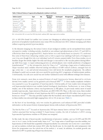
Table 2. Natural history of regenerative/dysplastic nodules in cirrhosis
Malignant Time (months) to HCC
Author (years) Type No Unchanged Disappeared
changes progression Mean (range)
[71]
Kondo et al. , 2011 LRN 17 - 13 (76%) 4 (24%) NA
Terasaky et al. [72] , 1998 LRN/DN 34 5 (15%) 4 (12%) 25 (73%) NA
Seki et al. [73] , 2000 DN 33 4 (9%) 14 (42%) 15 (49%) 18 (16-21)
Borzio et al. , 2003 LRN/DN 90 28 (31%) 44 (49%) 18 (20%) 22 (8-48)
[5]
Kobayashi et al. [74] , 2006 LRN/DN 154 29 (19%) 81 (53%) 44 (28%) NS
Iavarone et al. [76] , 2013 LRN/DN 36 11 (31%) 21 (53%) 4 (16%) 13 (7-27)
Sato et al. [75] , 2015 LRN/DN 92 19 (21%) 30 (32%) 43 (47%) NS
NA: not available; NS: not specified. DN: dysplastic nodules; LRN: large regenerative nodules
LG- or HG-DNs found that neither size increase nor changing on enhancing pattern emerged as accurate
predictors of neoplastic transformation since some DNs transformed into HCC without enlarging and others
without acquiring arterial hypervascularity.
In the dynamic imaging era, the natural history of pre-malignant nodules can be extrapolated from mainly
retrospective studies including nodules classified as non-enhancing/indeterminate at both CT and MRI to
differentiate them from progressed HCC. Studies before 2010 and mainly based on dynamic CT, confirmed
that some of these hypo-enhancing nodules carry a risk to acquire true radiologic hallmarks of malignancy
over time even though this risk at an individual level still remains unpredictable. From these studies, size at
baseline (larger the nodule, higher the risk) and changes in size and/or in the vascular pattern during follow-
up (shift from hypo- to hyper-enhancing pattern on arterial phase), were reliable predictors of malignant
[79]
transformation [77,78] . In the retrospective study by Chuo et al. including a large series of indeterminate
(hypo-vascular) small nodules observed during surveillance of HBV-related cirrhosis, emerged that old
age, initial size > 1 cm and arterial enhancement were risk factors for neoplastic progression. The authors
developed a useful and accurate risk score model for predicting HCC progression of indeterminate nodules.
Unfortunately, this risk score model was not further validated in series with different etiologic liver diseases.
From 2010 onwards, most data on natural history of small hypovascular lesions observed in cirrhosis
derived from studies carried out by gadoxeti-acid enhanced MRI [80-86] . In these studies neoplastic evolution
was assumed to occur by acquisition of hypervascularity. Unfortunately, all these studies were retrospective
and hypovascular/indeterminate nodules lacked histological classification. Furthermore, in most of these
studies, one of the inclusion criteria was hypointensity at HE-phase. As previously stated, most of small
nodules hypovascular , hypo-intense at He-phase, are eHCC/HG-DN. Thus, in the strict sense, these studied
focused on the natural history of early hypovascular HCCs or progressed HG-DN rather than on precursor
lesions as a whole [80-86] . This may partially explain why the transition rate to hyper-vascularized pHCC found
in these studies was extremely high ranging from 12% to 35% at one year [Table 3].
At the best of our knowledge, only two studies by gadoexetic-acid enhanced MRI provided reliable
information on the natural history of premalignant lesions with exclusion of hypovascular HCC.
[85]
The first by Kim et al. focused on hypovascular, HE hypo-intense lesions without T2W hyperintensity.
Authors assumed that having excluded T2 hyperintense nodules, the risk of inclusion of eHCC among
hypovascular hypointense nodules on HE phase was marginal.
In this study the rate of hypervascular transformation of precursor nodules, such as LG-DN and HG-DN
[86]
was 23 % at 3 years. In the second study, Sano et al. in a large series of small hypo-vascular nodules, iso/
hyperintense at HE found that acquisition of hypervascularity was extremely rare (0.6%) over 3 years follow-
up and no nodules evolved into mature HCC after the fourth year. In this study the only independent risk
factor for progression was the initial size of the nodule (> 10 mm). In addition, the nodule growth rate
showed 85% PPV in predicting of hypervascularization.