
Page 184 - Read Online
P. 184
Page 12 of 16 Borzio et al. Hepatoma Res 2019;5:15 I http://dx.doi.org/10.20517/2394-5079.2019.11
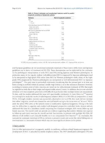
Table 4. Clinical, radiologic and morphologic features useful to predict
neoplastic evolution of dysplastic nodules
Features Risk of neoplastic evolution
Size
< 1 cm Low
> 1.5 cm High
Arterial enhancement at CT, MRI, CEUS
Hypo-vascularity Low
Hyper-vascularity High
HE phase at gadoxetate-enhanced MRI
Iso/hyper-intense Low
Hypo-intensity High
DWI
Hypo-intensity Low
Hyper-intensity High
Imaging features at follow-up
Stable size Low
Increasing size High
Stable vascular pattern Low
Acquired hyper-vascularity High
Sincronous HCC
No Low
Yes High
Histologic diagnosis
LG-DN Low
HG-DN high
LG-DN: low-grade dysplastic nodule; HG-DN: high-grade dysplastic nodule; HCC: hepatocellular carcinoma
and European guidelines do not recommend systematic treatment of these lesions while Asian and Japanese
guidelines are in favour of treatment od HG-DN. These discrepancies can be explained by the confusion
in the pathological interpretation of early HCC and DNs among Japanese and Western pathologists. In
particular, many of the vaguely nodular well-differentiated HCCs diagnosed by Japanese pathologists tend
to be interpreted as high-grade DNs rather than HCC by Western pathologists while, many of the high-
grade DNs diagnosed by Western pathologists are interpreted as well-differentiated HCC by Japanese
[87]
pathologists . This grey zone is particularly worrisome considering that the pursued goal is to treat any
lesion arising in cirrhosis within an optimal curable stage (within 2 cm as the maximum diameter). However,
according to western point of view, concerns are raised on the indiscriminate treatment of DNs that might
be regarded as futile due to their longer and unpredictable natural history. In addition, the accurate selection
of lesions with true neoplastic potential is still difficult in particular when multiple lesions are encountered.
To data, only few studies addressed this issue with questionable conclusions. In 2008, Kim et al. reported
[88]
in a retrospective study the results of radiofrequency ablation (RFA) of 21 HG-DNs as compared to 41 small
HCCs. Although complete necrosis was successfully obtained in 100 % of DN, this result did not translate
into either long-term overall and disease-free survival benefit owing to the occurrence of “de novo” HCCs
aside the initial DNs (48%) as the natural course of multicentric hepatocarcinogenesis. Owing to the lack
and the difficulties to organize and conduct well-designed prospective controlled trials, Korean authors
addressed this issue by a simulation model comparing two treatment strategies: RFA versus follow-up and
timely resection. This model could not provide any evidence supporting that nodular ablation was superior
to follow-up and timely resection for overall survival. Furthermore, in patients with multiple HG-DNs, RF
[89]
ablation of all nodule is not clinically feasible, as it can compromise liver function . In conclusion, the
rationale for systematic treatment of DN in cirrhosis at present is weak and carries the risk of falling into an
overtreatment, i.e., treatment of lesions which may not cause significant disease in the patient.
CONCLUSION
Like in other gastrointestinal oncogenetic models, in multistep cirrhosis-related hepatocarcinogenesis the
development of HCC is preceded by sizable dysplastic lesions. The IWP classification distinguish DN into