
Page 183 - Read Online
P. 183
Borzio et al. Hepatoma Res 2019;5:15 I http://dx.doi.org/10.20517/2394-5079.2019.11 Page 11 of 16
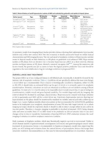
Table 3. Natural history of small hypovascular nodules at MRI and evaluated by gadoxetic acid uptake at hepatic phase
Author (years) Type HE intensity Nodules Acquaired Mean follow-up Risk factors
at baseline (n) hypervascularity (months)
Kumada et al. [81] , 2011 Retrospective Hypo- 49 6 (27%) 12 Size ≥ 15 mm
Motosugi et al. [80] , 2011 Retrospective Hypo- 135 16 (12%) Size ≥ 10 mm
Fat content
enlargement
Kim et al. [83] , 2012 Retrospective Hypo- 214 75 (35%) 11 Hyperintensity at DWI
Hyodo et al. [82] , 2013 Retrospective Hypo 160 50 (31%) 12 Rapid growth (tumor
volume doubling time
= 542 days)
T2W hyper-intensity
Higaki et al. [84] , 2014 Retrospective Hypo 60 10 (17%) 12 Higher growth rate
Kim et al. [85] , 2016 Retrospective Hypo 114 26 (23%) 42 T1w hyperintensity
No T2W Size > 10.5 mm
hyperintensity Previous HCC
Rapid growth rate
Sano et al. [86] , 2017 Retrospective Iso-hyper 663 6 (0.9) 36 Size > 10 mm
HCC: hepatocellular carcinoma
In summary, results from imaging-based studies provide evidence showing that indeterminate hypovascular
nodules may evolve into mature HCC but this transition is hardly predictable based on initial clinical
characteristics and MR imaging features. The risk and speed of neoplastic evolution of hypovascular nodules
seems to depend mostly on their behaviour at HE phase on gadoexetic-acid enhanced MRI. Hypo-intense
nodule at HE phase, have an elevated risk to became hypervascular pHCC in a short interval, whereas
nodules iso/hyper-intense at HE phase, showed a minimal oncogenic risk [80-86] . Among the numerous risk
factors found, the growth rate per se seems to have the highest positive predictive value and should be
regarded as the most reliable alarm ring for radiologic evolution to progressed HCC.
SURVEILLANCE AND TREATMENT
The proper follow-up of non-malignant lesions is still debated and, theoretically, it should be dictated by the
intrinsic risk of neoplastic evolution [Table 4]. Guidelines do not specifically address this issue even though
a strict follow up is recommended. Once a nodule is histologically classified as dysplastic, either LG-DN
or HG-DN, it should enter an enhanced follow-up the goal of which is to promptly capture its neoplastic
transformation. However, indications on such an enhanced surveillance are not uniform among different
guidelines. An interval of 3-4 months seems to be reasonable since it would ensure that, in case of malignant
transformation, nodule would not grow beyond curability. For nodule diagnosed only by imaging, the
interval should be dictated by radiologic characteristics. Hypovascularity coupled with hypo-intensity
at HE by gadoexetic-acid MRI call for a strict follow up. Conversely, hypovascular nodules showing iso/
hyperintensity at HE-phase should be monitored by standard six months interval and with a follow-up no
longer than 3 years. Eighteen months observation period as that recommended by AASLD/EASL guidelines
seems to be inadequate since neoplastic transformation of some DN may take longer interval. As to which
imaging technique should be employed, ideally it should be able to detect changes either in size and/or in
vascular profile. Therefore, a dynamic imaging is preferable to standard ultrasound. CEUS, being cheaper,
safer and more accessible than CT or MRI seems to be preferable in clinical practice since it can catch
changes either in size or vascularity. Gadoexetate-enhancing MRI with DWI evaluation remains the recall
imaging of reference to confirm neoplastic transition of nodules.
Early treatment of dysplastic nodules, which may theoretically improve survival is controversial. Unlike in
others human models of gastrointestinal carcinogenesis (colo-rectal and gastroesophageal cancer) where
treatment of precancerous lesions is recommended, in hepatocarcinogenetic model data supporting this
policy are still lacking and evidences from the natural history of preneoplastic lesions discourage their
systematic treatment. Recommendations on this issue by international guidelines are discordant. American