
Page 177 - Read Online
P. 177
Borzio et al. Hepatoma Res 2019;5:15 I http://dx.doi.org/10.20517/2394-5079.2019.11 Page 5 of 16
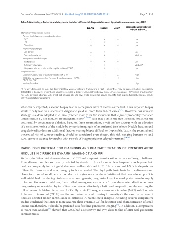
Table 1. Morphologic features and diagnostic tools for differential diagnosis between dysplastic nodules and early HCC
Diagnostic value between
LG-DN HG-DN eHCC HG-DN and eHCC
Elementary morphologic feature
Parenchimal changes, cytologic alterations
SCC - + + Low
LCC ± ± - Low
Clone like ± + + Low
Architectural changes
Cell density ± + + Low
Pseudoglands/acini - ± + Medium
Non-parenchymal changes
Portal tracts + + ± Low
Reticulin framework + + ± Medium*
Umpaired arteries or sinusoidal capillarization (CD34) ± ± + Low
Diagnostic tools
Stromal invasion/loss of ductular reaction (K7/19) - - ± High
HCC biomarkers expression (at least 2 markers among HSP70, - - -+ High
GPC3, GS, CHC)
Nodule-in-nodule - - ± High
*If frankly decreased or lost, the discriminatory value of reticulin framework is high. -: absent; ±: may be present but not necessarily
detectable in biopsy; +: present and usually detectable in biopsy. CHC: clathrin heavy chain; GPC3: glypican 3; HSP70: heat shock protein
70; LCC: large cell change; SSC: small cell change; LG-DN: low-grade dysplastic nodule; HG-DN: high-grade dysplastic nodule; eHCC:
early hepatocellular carcinoma
what can be expected, a second biopsy has the same probability of success as the first. Thus, repeated biopsy
would finally lead to a successful diagnostic yield in more than 90% of cases [8,21] . However, this invasive
strategy is seldom adopted in clinical practice mainly for the awareness that a priori probability that such
indeterminate 1-2 cm nodules are malignant is low [8,9,22,23] and that 2 cm is the size threshold to achieve the
best result by percutaneous ablation. Based on these assumptions, a wait and see strategy with the adoption
of a strict monitoring of the nodule by dynamic imaging is often preferred (see below). Nodule location and
coagulative disorders are additional features making biopsy difficult or impossible. Lastly, the potential and
theoretical risk of tumour seeding, should be considered even though, this risk, ranging between 1% and
2.7%, seems to balance favourably with the risk of inappropriate or delayed treatment [24-26] .
RADIOLOGIC CRITERIA FOR DIAGNOSIS AND CHARACTERIZATION OF PRENEOPLASTIC
NODULES IN CIRRHOSIS DYNAMIC IMAGING CT AND MRI
To date, the differential diagnosis between eHCC and dysplastic nodules still remains a radiologic challenge.
Premalignant nodules are usually detected by standard US as hypo- or, less frequently, as hyper-echoic
nodules completely indistinguishable from well-established HCC. Thus, standard US is inadequate for
differential diagnosis and other imaging tools are needed. The physiopathologic basis for the diagnosis and
characterization of small hepatic nodules by imaging rests on characteristics of their vascular supply. It is
well established that during cirrhosis-related oncogenesis, progressive loss of normal portal vascular supply
in favour of increase arterial one, the so called neoangiogenesis, occurs. This nodular arterialization becomes
progressively more evident by transition from regenerative to dysplastic and neoplastic nodules reaching the
full expression in high-differentiated HCCs. Dynamic CT, magnetic resonance imaging (MRI) and Contrast-
Enhanced Ultrasound (CEUS) are the contrast-enhanced imaging to investigate the vascular pattern of
nodules detected under surveillance in cirrhosis. A recent meta-analysis including several comparative
studies confirmed that MRI is more accurate than dynamic CT for detection and characterization of small
[27]
lesions and therefore, it should be preferred as a first line panoramic imaging . In addition, a comparative
[28]
13-years meta-analysis showed that CEUS had a sensitivity and PPV close to that of MRI with gadoexetic
contrast media.