
Page 214 - Read Online
P. 214
Giakoustidis et al. Sorafenib-everolimus for metastases after liver-transplantation
liver cirrhosis due to ALD. He was presenting portal and 8th left ribs. Magnetic resonance imaging (MRI)
hypertension, ascites and episodes of encephalopathy. scan failed to reveal any additional findings. Therapy
His model for end-stage liver disease (MELD) remained the same.
score was 21. He was transplanted with piggy-back
technique, from a heart-beating donor. Cold ischemia Another 99mTc-HDP bone scanning 18 months post LT
time was 9 h. He was put on triple immunosuppression showed, for the first time, regression of the rib lesions,
maintenance therapy with prednizolone, mycophenate while the known 2 spinal lesions were significantly
mofetil and cyclosporine. minimized. Therapy remained unaltered. Patient’s
clinical condition was excellent.
The explant’s pathology report revealed the presence
of two incidental HCC lesions measuring 15 and 20 mm, Finally, 28 months post LT, a new bone scanning certified
with no portal involvement, of medium differentiation, the complete regression of all the osteolytic lesions
with pseudocapsule, clear-cell type, without extrahepatic [Figure 2].
nodules or other findings. The post-operative course
was uneventful. His immunosuppression therapy was DISCUSSION
changed to tacrolimus and everolimus, along with
tapering of prednizolone. Tacrolimus and everolimus HCC is the third cause of cancer related mortality
levels were monitored. nowadays, according to World Health Organization
(WHO). The primary etiologic factor is liver cirrhosis.
Two months post transplantation the patient complained To the present case, HCC was incidental finding
of back pain. Bone Scanning 99m Technetium in the explant. A prior transplantation computed
helix destabilizing protein (99mTc-HDP) revealed tomography (CT) failed to detect the presence of liver
2 osteoblastic lesions on the T8 and T11, possibly or extrahepatic lesions. Additionally, AFP levels were
secondary-HCC lesions. Prednizolone was ceased and
sorafenib 400 mg bid was initiated, along with ibandronic low [Table 1], failing to justify a position emission
acid (diphosphonic acid) qd. Radiotherapy was induced, tomography (PET) scan preoperatively. Even if
photons 60Co. He received a total of 2,300 centigray the patient was evaluated for Milan Criteria (MC),
(cGy), in doses of 46 cGy, 5 times/week. according to the explants’ pathology, the patient would
be inside MC. Moreover, piggy-back technique is the
Otherwise the patient was in good condition. His standard LT procedure performed by our center, like
kidney function with radioisotope renography with many other centers universally. It does not consisting
99m Technetium diethylene triamine pentoacetic a risk factor for HCC recurrence, compared to the
acid (99mTc-DTPA) was 52 mL/min/1.73 m . Alpha classic technique.
2
fetoprotein (AFP) level was 6.6 ng/mL.
The induction of sorafenib, an oral multi-kinase
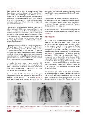
Seven months after the first discovery of the spinal inhibitor, targeting HCC control, demands compensated
osteoblastic lesions, the repetition of the 99mTc-HDP, liver function, and applies to patients with advanced
[4]
revealed further progress of the disease [Figure 1]. HCC. Regarding HCC recurrence post LT, the current
New lesions were being detected at the 5th, 6th, 7th, strategy remains controversial. Recurrence can be
Planar Planar Planar
POST ANT LAO
Figure 1: Initial 99m Technetium helix destabilizing protein scintigraphy showing metastatic lesions in the spine and ribs. POST:
posterior; ANT: anterior; LAO: left anterior oblique
206 Hepatoma Research ¦ Volume 3 ¦ September 20, 2017