
Page 104 - Read Online
P. 104
Link et al. Roles of p53 in extrinsic factor-induced liver carcinogenesis
INTRODUCTION probability of being diagnosed at advanced stages, as
well as poor responses to systematic chemotherapy
Liver cancer is the 6th most common cancer in and radiation therapy, prognosis of HCC is particularly
men and the 9th most common cancer in women bleak with an incidence to mortality ratio of 0.95 and a
with the 3rd highest mortality rate of all cancers 5-year survival rate around 17.5%. [2,13]
globally. [1,2] The majority of these cases (about 80%)
occur in Eastern Asia, South-Eastern Asia, Mid- Molecular mechanisms involved in liver carcinogenesis
Africa, and West Africa, within the context of viral remain unclear. The tumor suppressor p53, a
hepatitis. [2-4] Although there are genetic etiologies for transcription factor that regulates many downstream
hepatocellular carcinoma (HCC) including hereditary target genes regulating cell cycle progression,
hemochromatosis and α1-antitrypsin deficiency, [5-7] a p o pto s is , D N A r ep a i r, sen e s c en c e, a nd
viral hepatitis, as well as exposure to other extrinsic metabolism, [14,15] is one of the most commonly mutated
factors, such as aflatoxin B1 (AFB1), polyvinyl chloride genes in HCC. [16,17] Indeed, p53 is the most commonly
(PVC), a poor diet inducing non-alcoholic fatty liver mutated human gene, occurring in > 50% of all human
disease (NAFLD), and excess iron exposure, remain cancers. [18] Additionally, in some HCC cases, proteins
among the most common causes of liver cancer. [8,9] such as a 26S proteasome regulatory protein, gankyrin,
Despite vaccinations for hepatitis B virus (HBV), new and a p53-specific ubiquitin ligase, murine double
treatments for hepatitis C virus (HCV), regulations minute 2 (MDM2), are elevated, hence decreasing
governing PVC production, and restrictions preventing p53 protein levels. [19,20] MicroRNAs (miRNAs) can
AFB1 contamination of food products, countries still also inhibit p53 activity; specifically, miRNA-24, when
struggle to prevent liver cancer. [9,10] dysregulated in HCC, is shown to promote invasion
and metastasis by decreasing p53 levels. [21] Thus, p53
Surgical resection is currently the preferred treatment, activity is impaired by multiple mechanisms in HCC,
and liver transplantation is ultimately the most effective hence contributing to HCC genesis. In this review
therapeutic modality of HCC; however, it is limited by article, we focus on HCC-inducing extrinsic factors that
the availability of suitable organs. [11,12] Due to a high are etiologically associated with p53 [Table 1].
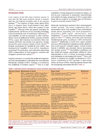
Table 1: Extrinsic factors causing liver cancer and their association with p53
Extrinsic factors Mechanisms of action Roles of p53 References
AFB1 AFB1 is metabolized to AFB1-8,9-epoxide to AFB1 frequently causes p53 R249S mutation [25,29,34]
7
form AFB1-N -guanine adducts, leading to which enhances IGF-2 expression
specific mutation at p53 codon 249 (p53 R249S )
VC VC activated by CYP2E1 is converted into It is unclear whether p53 plays protective [41,43,44]
chloroethylene oxide, which forms bulky DNA roles in VC-induced liver cancer
adducts, leading to A>T transversions in the
genome
NAFLD NAFLD-induced hepatitis leads to cirrhosis and The miR-34-SIRT1-p53 pathway plays a role [49,51-57]
HCC, and dysregulation of NF-kB signaling, the in the progression of NAFLD. However, the
Pl3K-ATK-PTEN pathway, insulin resistance, and direct role of p53 in the NAFLD-mediated
expression of certain miRNAs (e.g. miR-34) is HCC is unknown
suggested; however, the molecular mechanisms
behind NAFLD-mediated HCC remain unclear
Iron Excess iron generates ROS and decreases p53 Chronic iron overload reduces p53 protein [64,68-70]
activity, leading to HCC genesis levels by heme-mediated degradation or
increased MDM2 levels, which can increase
intracellular iron levels via a decrease
in ISCUC2, thus further promoting HCC
development
HBV HBV-induced HCC occurs following repeated Although direct involvement of p53 in [76,81,82,85,99,100]
inflammation-liver regeneration-cirrhosis process, HBV-induced HCC is unclear, functional
as well as through oncogenic function of HBx and inactivation of p53 by HBx and Ct-HBx may
Ct-HBx in both p53-dependent and -independent contribute to HCC progression
manners
HCV The majority of HCV-mediated HCC is via There is no direct evidence showing [112,118,119,123-
cirrhosis. But HCV core protein, NS3, and NS5 dependency of HCV-induced HCC on p53. 125,131,132]
are implicated in HCC development in both p53- However, HCV core protein, NS3, and NS5A
dependent and -independent manners inhibit p53 activity by binding to p53, altering
subcellular localization, or modulating post-
translational modifications
AFB1: aflatoxin B1; VC: vinyl chloride; NAFLD: non-alcoholic fatty liver disease; HBV: hepatitis B virus; HCV: hepatitis C virus; HCC:
hepatocellular carcinoma; ROS: reactive oxygen species; MDM2: murine double minute 2; ISCUC2: iron-sulfur cluster enzyme 2; Ct-HBx:
HBx variants with C-terminal truncations
96 Hepatoma Research ¦ Volume 3 ¦ June 6, 2017